Parasagittal and Parafalcine Meningiomas: What Are They?
Tumors known as meningiomas develop from the meninges, which are the protective coverings that envelop the brain and spinal cord. Although the majority of meningiomas are benign, the pressure they place on other brain structures can result in serious issues.
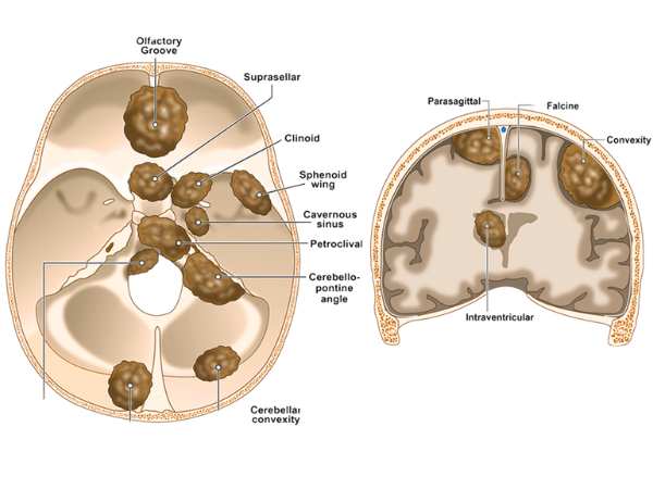
Two major forms of meningiomas that develop along the brain’s midline are parasagittal and parafalcine:
- Parasagittal Meningiomas: Develop close to the major vein that runs along the top of the brain, the superior sagittal sinus.
- The parafalcine The falx cerebri, a sickle-shaped fold of dura mater that divides the two cerebral hemispheres, is where meningiomas form.
Because of their position, these Tumors may induce symptoms even when they develop slowly, particularly if they compress vital brain regions or venous sinuses.
Reasons & Risks
Meningiomas are linked to a number of risk factors, while their exact cause is unknown:
- Genetic susceptibility (e.g., type 2 neurofibromatosis)
- Exposure to radiation
- More prevalent in women, hormonal impacts
- Usually diagnosed between the ages of 40 and 70
Parasagittal/Parafalcine Meningioma Symptoms
The tumor’s location and size affect the symptoms. These Tumors frequently impact the motor and sensory parts of the legs because they develop close to the midline of the brain. Typical signs and symptoms include:
- One or both legs feeling weak or numb
- Seizures
- Headache
- Changes in cognition or personality
- Abdominal control loss (in big Tumors)
- Indications of elevated intracranial pressure (when there is brain swelling or sinus compression)
Diagnosis
A thorough diagnosis consists of:
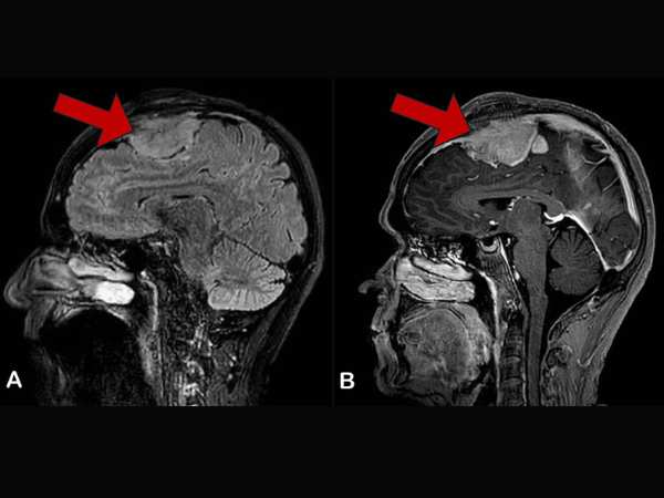
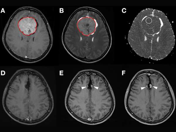
- The gold standard for identifying and describing meningiomas is MRI with contrast.
- A CT scan can be useful in detecting any calcifications or involvement of the bones.
- MR Venography: Crucial for assessing the superior sagittal sinus’s involvement
- To confirm the diagnosis and classify the Tumor as benign, atypical, or malignant, a biopsy may be required.
Options for Treatment
Tumor size, location, growth rate, and symptoms all impact treatment options:
- Observation
Regular imaging follow-up may be advised for older patients with tiny, asymptomatic Tumors.
- Excision via Surgery
The primary treatment for accessible Tumors that are producing symptoms is surgery. However, because parasagittal/parafalcine meningiomas are close to important blood arteries like the superior sagittal sinus, they can be difficult to fully remove.
- Radiosurgery Using Gamma Knife
This is a very successful non-invasive procedure that can be utilised either:
- As the main course of treatment for minor Tumors
- Targeting any remaining Tumors after surgery
- Regarding recurring Tumors
Radiosurgery with Gamma Knife for Parasagittal/Parafalcine Meningiomas
Gamma Knife Benefits for These Tumors:
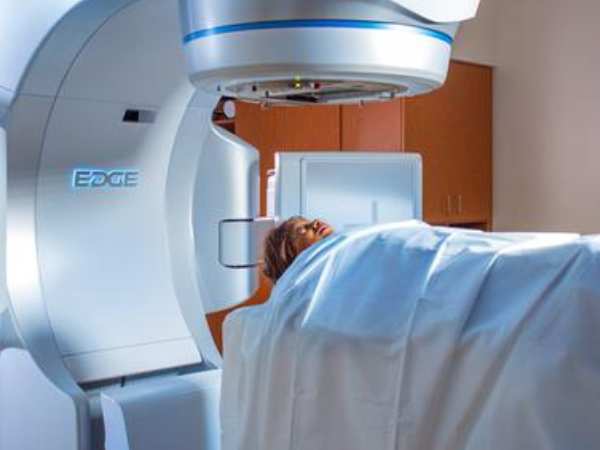
- Non-invasive; no anaesthesia or incision is necessary
- Quick recovery from an outpatient procedure
- Perfect for Tumors that are close to important structures (such as the superior sagittal sinus).
- Low rates of complications
- Elevated rates of Tumor control (>90% in benign meningiomas)
A Candidate Is Who?
- Individuals with Tumors that are small to larger in size
- Tumors near or involving the venous sinuses, where there is a greater risk of surgery
- Patients who are not surgically fit
- Recurring Tumors following previous operations
Overview of the Procedure:
- Planning based on MRI
- For accuracy, a lightweight frame or frameless mask is utilised.
- Usually, one session is enough to finish the treatment.
- On the same day, patients go home.
Prognosis and Follow-Up
The long-term results of parasagittal/parafalcine meningiomas are quite good when they are treated with surgery, Gamma Knife, or both. To check for Tumor control or recurrence, routine MRI scans are required.
In conclusion
Despite usually being benign, parasagittal and parafalcine meningiomas can have a major effect on neurological function because of where they are located. Excellent results with little risk can be achieved with early diagnosis and customised treatment, which frequently combines microsurgery and Gamma Knife Resonance Imaging.
Source:
- https://pmc.ncbi.nlm.nih.gov/articles/PMC6889965/
- https://www.sciencedirect.com/science/article/abs/pii/B9780128221983000318
- https://www.healthline.com/health/brain-tumor/parafalcine-meningioma
- https://www.tandfonline.com/doi/full/10.1080/02688697.2019.1635988?scroll=top&needAccess=true
- https://www.sciencedirect.com/science/article/abs/pii/S1878875018329401
- https://www.medicalnewstoday.com/articles/parasagittal-meningioma#diagnosis
- https://pubmed.ncbi.nlm.nih.gov/26115473/
- https://pubmed.ncbi.nlm.nih.gov/23930861/
- https://www.redjournal.org/article/S0360-3016(09)01590-9/fulltext