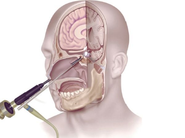
Endoscopic Endonasal Transsphenoidal Approach for Pituitary Adenoma
What is a Pituitary Tumor?
Unusual growths that form in the pituitary gland are known as pituitary tumours. The size of this gland looks similar to that of a pea. It is situated at the base of the brain, behind the nose. The pituitary gland produces too many hormones that regulate vital bodily processes as a result of certain of these tumours. Others may result in insufficient production of those hormones by the pituitary gland.
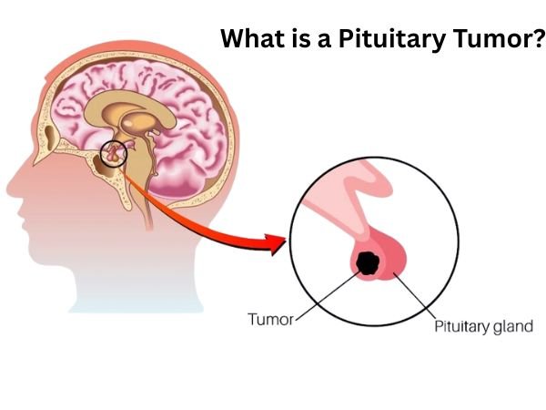
The majority of pituitary tumours are harmless. Thus, they are not cancer. Pituitary adenomas are another term for these benign tumours. The majority of adenomas grow slowly and remain in the pituitary gland or the tissue around it. Usually, they don’t spread to other bodily parts.
Treatment options for pituitary tumours are numerous. Surgery may be used to remove the tumour. Alternatively, radiation therapy or medication may be used to restrict its growth. Medicine is sometimes used to control hormone levels. A combination of these therapies may be recommended by the doctor. Sometimes the best course of action is observation, sometimes known as a “wait-and-see” strategy.
Through the nose and sphenoid sinus, the pituitary gland and surrounding tissues can be accessed using the endonasal transsphenoidal method, a minimally invasive surgical technique. This method is frequently used to treat disorders like acromegaly or Cushing’s disease, remove pituitary tumours, and correct other anomalies in the base of the skull.
Despite being benign, pituitary adenomas can have a significant impact on a patient’s health, resulting in everything from hormone abnormalities to vision impairment via optic chiasm compression. Pituitary surgery has been changed by the endoscopic endonasal transsphenoidal approach (EETA), which provides a less invasive method of tumour removal with fewer side effects and a quicker recovery.
Endoscopic endonasal surgery: What is it?

A cutting-edge technique called endoscopic endonasal surgery removes the need for open surgery by entering the nasal passages and accessing the pituitary gland and surrounding structures. This method enables surgeons to precisely view and access the surgical area by using a high-definition endoscope, a tiny, flexible tube equipped with a light and camera.
The endonasal technique removes brain tumours, numerous midline skull base tumours, and pituitary adenomas by using the nostrils as natural surgical passageways.
For practically all pituitary adenomas, Rathke’s cleft cysts, the majority of craniopharyngiomas, clival chordomas, and numerous midline menigiomas, the endonasal route is the recommended surgical technique. In skilled hands, the endonasal approach has become a safe and successful treatment that uses high-definition endoscopy (a surgical telescope), surgical navigation (sometimes known as “GPS for the brain”), and Doppler ultrasound for carotid artery localisation.
The main benefits of the endonasal approach:
- Avoiding brain retraction
- Requiring little to no treatment of the optic nerves
- Avoiding facial incisions
- Rarely requiring nasal packing
Endoscopic Endonasal Surgery: Advantages
When treating pituitary disorders, endoscopic endonasal surgery has a number of advantages over standard techniques.
These consist of:
- Minimally invasive: By performing the entire procedure through the nose, no exterior cuts are made, and the surrounding tissues are not as severely damaged.
- Faster recovery: In comparison to standard surgery, patients frequently report shorter hospital stays (48–72 hours post-surgery), less pain, and a quicker recovery in general.
- Better visualisation: The endoscope makes it easier to see the surgical site, which enables more accurate tumour removal and lowers the risk to adjacent vital tissues.
- Decreased risk of problems: Endoscopic endonasal surgery considerably reduces the risk of complications including infection and CSF leakage.
Knowing How to Use the Endoscopic Endonasal Method

In the past, microscopic transsphenoidal surgery or open craniotomy were used to access pituitary adenomas. However, direct visibility and improved mobility have been made possible by the development of endoscopic procedures, which has improved surgical results.
Using a high-definition endoscope, EETA involves getting into the pituitary gland through the sphenoid sinus and nostrils. By doing away with the necessity for external incisions, this method reduces post-operative morbidity and stress to the surrounding tissues.
The Preoperative Assessment of Endoscopic Endonasal Surgery
- Patients will have a thorough preoperative evaluation prior to endoscopic endonasal surgery, which may involve hormonal testing, imaging studies, and consultations with our experienced neurosurgeons and endocrinologists. This comprehensive evaluation guarantees personalised treatment programs targeted to the unique requirements of every patient.
Method of Surgery
- The patient is put under general anaesthesia for the procedure. With the help of an otolaryngologist, the neurosurgeon inserts the endoscope through the nostrils to reach the pituitary gland and its surroundings. While maintaining normal pituitary gland function, advanced tools and procedures are used to remove or treat the pituitary tumour or other underlying problems.
Recovery and Postoperative Care
- Before being moved to a hospital room after endoscopic endonasal surgery, patients are closely watched in the recovery area. Experienced medical staff offers complete postoperative care, including pain management, hormone level monitoring, and handling any possible complications. The majority of patients recover more easily and can resume their regular activities in 2-3 weeks.
- Patients are kept under observation for diabetes insipidus and hormone abnormalities, which may necessitate short-term hormone replacement treatment. Mild headaches and nasal congestion are frequent but go away in a few weeks.
The Surgical Procedure in Steps
- Patient Preparation: In order to schedule surgery, patients get CT and MRI scans. Vasoconstrictors are used to clear congestion in the nose in order to reduce bleeding.
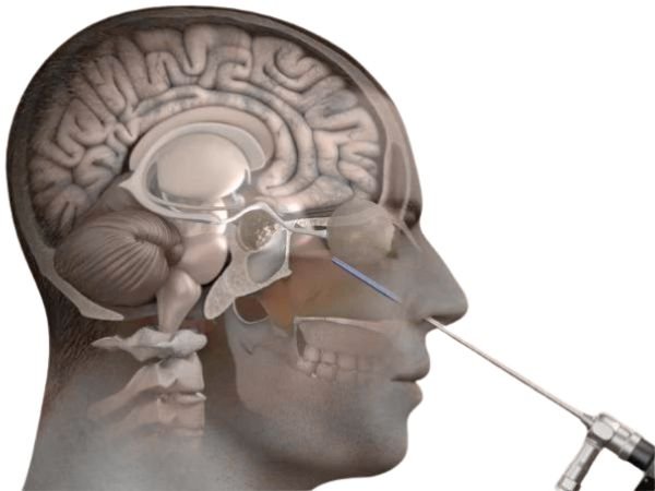
- Nasal Entry: To view the sphenoid sinus, a rigid endoscope, typically 4 mm in diameter, is placed via one nostril.
- Sphenoidotomy: The sella turcica is revealed by delicately opening the sphenoid sinus’s anterior wall.
- Tumour Removal and Dural Opening: Using microsurgical tools, the adenoma is carefully removed once the dura is cut.
- Reconstruction: To stop cerebrospinal fluid (CSF) leakage, the sellar floor is reconstructed using synthetic materials or autologous tissue grafts.
Pituitary adenomas that can be accessed with the endoscopic endonasal approach include:
Pituitary adenomas including:
- Acromegaly
- Crushing’s Disease
- Prolactinomas
- TSH-Secreting Adenomas
- Non-Functional Adenomas
- Pituitary Apoplexy
- Recurrent Adenomas
Non-Pituitary Tumors
- Craniopharyngioma
- Rathke’s Cleft Cyst
- Sellar Arachnoid Cyst
- Meningioma (tuberculum sellae, parasellar, cavernous sinus regions)
- Clival Chordoma
- Sinonasal Carcinoma
- Olfactory Neuroblastoma
Not all midline tumours can be approached using the endonasal technique. A different minimally invasive procedure, like a standard craniotomy or a supraorbital eyebrow craniotomy, might be suggested for certain patients.
Potential Challenges and Considerations
- Despite being a major breakthrough, EETA requires a highly qualified surgical team. Important difficulties include:
- CSF leak risk, which calls for careful dural closure
- Possibility of incomplete tumour removal, especially in cases of invasive macroadenomas
- Expert rhinologists are required to handle the complex nasal structure.
In conclusion
In the treatment of pituitary adenoma, the endoscopic endonasal transsphenoidal technique represents a paradigm change. It provides a great substitute for traditional approaches due to its less invasive nature, enhanced visualisation, and quicker recovery. This method keeps developing as technology does, improving surgical accuracy and patient outcomes even further.
Source:
- https://www.yalemedicine.org/clinical-keywords/endonasal-transsphenoidal-approach#:~:text=Definition,the%20nose%20and%20sphenoid%20sinus.
- https://pubmed.ncbi.nlm.nih.gov/21181278/
- https://www.sciencedirect.com/science/article/pii/S2214751921000487
- https://www.frontiersin.org/journals/oncology/articles/10.3389/fonc.2021.643550/full
- https://www.pacificneuroscienceinstitute.org/pituitary-disorders/treatment/endoscopic-endonasal-surgery/#:~:text=The%20endonasal%20route%20is%20the,chordomas%20and%20many%20midline%20menigiomas.
- https://www.mayoclinic.org/diseases-conditions/pituitary-tumors/symptoms-causes/syc-20350548
- https://www.cancer.gov/publications/dictionaries/cancer-terms/def/pituitary-tumor
- https://www.ninds.nih.gov/health-information/disorders/pituitary-tumors