

When a tumour, epilepsy-related condition, or other brain lesion is situated near regions of the brain involved in speech, movement, feeling, vision, memory, or comprehension, brain surgery demands extraordinary precision.
Thankfully, neurosurgery nowadays is much more sophisticated than just choosing where to operate based on a brain scan. These days, neurosurgeons may identify key functional areas and design surgery around them using brain mapping, functional MRI, tractography, neurophysiological monitoring, and intraoperative stimulation.
The objective is straightforward:
As much of the aberrant tissue can be safely removed or treated while maintaining the patient’s most important functions.
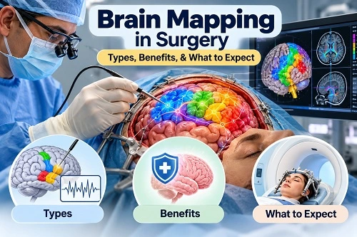
This strategy is especially crucial when eloquent brain regions, areas of the brain that regulate vital processes like speaking and movement are involved in surgery.
Brain Mapping: What Is It?
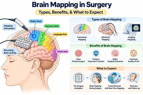
A set of methods known as “brain mapping” are used to determine which parts of a patient’s brain are involved in critical processes like:
- Language and speech
- Motion, Sensation, Vision, and Memory
- Recognising and thinking
Everybody’s brain is not structured the same way. Additionally, functional areas may occasionally move or reorganise due to tumours and other brain disorders.
As a result, a brain scan that reveals the location of a tumour may not necessarily provide the surgeon with precise information about the patient’s speech or motor abilities.
The neurosurgical team can arrange a safer procedure with the additional knowledge provided by brain mapping.
Why Is Brain Mapping Crucial Prior to Brain Surgery?
Certain brain tumours are situated in close proximity to regions that perform vital activities.
For instance:
- Movement of the arm, hand, face, or leg may be impacted by a tumour close to the motor cortex.
- Speaking, comprehending, reading, or naming may all be impacted by a tumour close to language-related regions.
- Communication between several brain regions may be impacted by a lesion near significant white-matter pathways.
The neurosurgeon’s task is to achieve a balance between two objectives:
- As much aberrant tissue should be removed as safely as feasible.
- Preserve the neurological function of the patient.
Prior to and during surgery, functional mapping can assist in identifying deeper white-matter networks and critical cortical regions.
How Does Brain Mapping Take Place?
Depending on the patient’s condition and the lesion’s location, brain mapping may be done prior to, during, or both procedures.
1. fMRI, or functional MRI
A non-invasive imaging examination called a functional MRI examines variations in blood flow linked to brain activity.
The patient may be required to complete particular tasks during an fMRI, such as:
- Talking
- Giving items names
- Using a foot or hand
- Finger tapping
- Completing specific tasks related to language
The brain areas that become active during these tasks can be identified with the aid of the generated photographs.
Therefore, fMRI can offer valuable information for surgical planning, especially if a tumour is near language or motor areas.
2. Tractography and Diffusion Tensor Imaging (DTI)
The surface of the brain does not regulate every significant brain function.
Millions of nerve fibres connect various parts of the brain. We refer to these channels as white-matter tracts.
The direction of water transport inside white matter can be investigated using Diffusion Tensor Imaging (DTI). Tractography, a representation of significant nerve-fiber routes, can then be produced by specialised computer processing.
This could aid surgeons in comprehending the connection between a tumour and significant circuits involved in:
- Language
- Movement
- Vision
- Sensation
Surgeons can organise the surgical approach around significant functional structures with the aid of preoperative functional imaging and tractography.
An Awake Craniotomy: What Is It?
A specific type of brain surgery called an awake craniotomy involves the patient being conscious for a portion of the procedure.
Without pain management, the patient is not expected to undergo the surgery. Medication is utilised to preserve comfort and lessen anxiety, and the scalp is gently numbed. The patient may be awake when neurological testing is necessary and asleep throughout other parts of the procedure.
In the mapping phase, the patient might be requested to:
- Speak
- Count
- Identify images or items
- Go through/Read
- Respond to queries
- Lift your arm or leg
- Execute basic duties
The neurosurgical team examines the patient’s reactions concurrently. This enables the surgeon to evaluate brain activity in real time.
Why Would Brain Surgery Require a Patient to Be Aware?
Being awake for some parts of operation might yield important information, despite the fact that this may sound frightening.
Let’s say a tumour is situated close to a speech-related area.
Imaging may indicate the location of the language-related region prior to surgery. The surgeon could, however, require immediate confirmation of the areas that are critical for that particular patient.
The patient may be asked to identify things while particular areas are activated during awake mapping.
The surgical team is informed that the stimulated area is functionally significant if it momentarily impairs speaking or naming.
By asking the patient to carry out particular motions, the same idea can be applied to motor functions.
The surgical team can safeguard critical cerebral functions with the use of this real-time input.
How Is Speech Protected by Brain Mapping?
Multiple brain areas and related pathways are involved in the complicated function of language.
Important language-related functions may include the following, depending on the individual:
- Making a speech
- Learning a language
- Giving items names
- Reading about
- Composing
- Repetition of words
- Language processing
The patient may be asked to read words, name objects, identify photographs, or complete other language activities during awake language mapping.
The surgical team watches to see if stimulation makes the task momentarily difficult.
This data aids in determining which regions need to be shielded during tumour excision.
How Is Movement Protected by Brain Mapping?
Additionally, a network that connects neural pathways and the motor regions of the brain controls movement.
Stimulation may aid in the identification of regions involved in the movement of:
- Face
- Arm
- Hand
- Leg
When appropriate, the patient may also be instructed to move a specific bodily part.
The information can assist the surgeon in locating critical motor structures and avoiding them as much as possible when doing surgery.
What Takes Place If the Tumour Is Extremely Near a Vital Brain Region?
One of the most significant circumstances in which functional mapping can be helpful is this one.
The surgeon may decide that removing all of the tumor’s visible parts could pose an intolerable risk to the nervous system if the tumour is in a crucial functional area.
Maximum safe resection could be preferable to trying removal at any costs in certain circumstances.
The precise surgical objective is determined by:
- Type of tumour
- Location of the tumour
- Size of the tumour
- Connection to brain tissue that functions
- Connection to significant white-matter pathways
- The neurological state of the patient
- Treatment options that are available
The goal of contemporary glioma surgery is to preserve cerebral function while minimising the amount of tumour removal.
Is Brain Mapping Limited to Brain Tumours?
No.
For some disorders, brain mapping may be an option if safe surgery requires the identification of key functional brain regions.
Applications could include the following, depending on the clinical setting:
- Brain cancers
- Gliomas
- Surgery for epilepsy
- Lesions close to language areas
- Lesions close to motor regions
- A few other neurosurgical techniques
The location of the anomaly and the functions that may be at risk determine how it is used.
Before Surgery Brain Mapping vs. During Surgery Brain Mapping
| Feature | Before Surgery | During Surgery |
|---|---|---|
| Main Purpose | Surgical Planning | Real-time functional assessment |
| Common Techniques | fMRI, DTI/tractography, nTMS, and other tests | Direct cortical/sub-cortical stimulation, neurophysiological monitoring |
| Patient Awake? | Usually yes | Maybe awake during selected portions |
| Information Obtained | Estimated location of functional area/pathways | Functional response during the operation |
| Main Benefit | Helps plan the surgical approach | Helps guide surgical limits in real time |
Frequently, these methods are combined instead of depending on just one test.
Can Brain Mapping Ensure the Preservation of Speech or Movement?
No.
Utilizing brain mapping is crucial in minimizing surgical risks, yet it does not assure that a patient will maintain full neurological function post-surgery.
The brain is extremely intricate. A tumor might invade or shift crucial areas, and some functions rely on interconnected networks rather than a single specific location.
Various other elements can impact the result, such as:
- Location of the tumor
- Nature of the tumor
- Size and extent of infiltration
- Existing neurological impairments
- Surgical intricacy
- Swelling post-surgery
- Bleeding or other issues
- Unique variations in brain structure
Hence, brain mapping should be seen as a method to reduce risks and plan surgeries, rather than a guarantee of a specific result.
Is Awake Brain Surgery Painful?
The brain itself lacks pain receptors unlike the scalp and other tissues.
During awake craniotomy, local anesthesia and medications are carefully administered to the scalp and surgical area. The patient might be conscious during the functional testing phase but is ensured comfort by the anesthesia team.
Patients might feel sensations like pressure, movement, sounds, or tugging instead of intense pain.
The patient’s comfort and status are continuously monitored by the anesthesia and neurosurgical teams.
What Does the Patient Experience During Brain Mapping?
Each patient’s encounter is unique.
While undergoing brain mapping, the patient may be interrogated with simple questions or given tasks like:
- “What is this item?”
- “Can you count from one to ten?”
- “Can you move your hand?”
The tasks are generally straightforward and recognizable.
The objective is not to evaluate intelligence but to determine if specific brain regions are engaged in specific functions.
Who Determines Whether Brain Mapping Is Necessary?
The decision is tailored to the individual.
Depending on the case, the neurosurgeon, neurologist, neuropsychologist, neuroradiologist, anesthesiologist, and other team members may collaborate on the planning.
Brain mapping might be particularly contemplated when a lesion is near an area that controls a crucial neurological function.
Not all brain surgeries necessitate awake mapping.
Before undergoing surgery, various evaluations are conducted by the medical team:
- Neurological assessment: The physician examines muscle strength, coordination, sensory perception, verbal communication, linguistic ability, eyesight, and other neurological capabilities.
- Brain scans: Imaging techniques like MRI or CT scans are utilized to determine the size and position of the abnormality.
- Functional imaging: In specific cases, examinations such as functional MRI or DTI-based tractography might be carried out.
- Neuropsychological or linguistic evaluation: When needed, a specialist evaluates speech, language, memory, focus, and other cognitive functions.
These assessments establish a starting point for comparing post-surgery functionality.
What Happens After Surgery?
After the surgical procedure, the patient is carefully monitored to understand what to expect before and after brain surgery.
The medical team reviews:
- Verbal communication
- Muscle strength
- Sensory perception
- Coordination
- Eyesight
- Memory and cognitive abilities
- Other neurological functions
Temporary weakness or speech issues may arise in some patients due to swelling or surgical intervention. The recovery process is dependent on each individual case, including understanding life after brain tumour surgery.
If necessary, rehabilitation like speech therapy, physical therapy, occupational therapy, or cognitive rehabilitation may be suggested.
What Are the Advantages of Brain Mapping?
Benefits of brain mapping may include:
- Enhanced comprehension of individual brain activity: Functional regions can differ among people.
- Enhanced surgical preparation: Preoperative mapping aids the surgical team in understanding how neurosurgeons learn brain anatomy in correlation between a lesion and vital functional areas.
- Immediate data: Intraoperative mapping can offer instant feedback during specific surgeries.
- Safeguarding crucial neurological functions: Mapping can assist in recognizing areas that need to be conserved.
- Assistance for achieving maximum safe removal of tumors: Functional information, when suitable, can aid surgeons in balancing tumor elimination with the preservation of neurological function.
Are There Dangers Linked to Brain Mapping?
Brain mapping is conducted as part of a meticulously planned neurosurgical procedure, but the overall operation still involves risks.
Potential risks can include:
- Temporary or permanent muscle weakness
- Difficulties with speech or language
- Seizures
- Bleeding
- Infection
- Swelling of the brain
- Complications related to anesthesia
- Alterations in memory or cognitive abilities
The specific risks can vary significantly from one patient to another.
Your neurosurgeon should outline the individual risks and anticipated benefits before the surgery.
Brain Mapping and the Future of Precise Neurosurgery
Modern neurosurgery is progressively moving towards tailored and precision-guided treatment.
Instead of treating every patient’s brain as anatomically the same, surgeons can integrate:
- High-definition MRI
- Functional MRI
- DTI and tractography
- Neuronavigation
- Neurophysiological monitoring
- Cortical and subcortical stimulation
- Conscious language or motor evaluations
- Intraoperative imaging
To create a customized surgical approach for the patient.
Research is ongoing to enhance how these technologies are integrated and how functional results are measured. Recent interdisciplinary suggestions have also highlighted the standardization of mapping, monitoring, and intraoperative decision-making in glioma surgery.
When is the Right Time to Discuss Brain Mapping with a Neurosurgeon?
In the event of a brain tumor diagnosis or another brain issue situated close to crucial areas controlling speech, movement, vision, or other essential functions, it is advisable to raise the following questions with the neurosurgical team:
- How close is the lesion to a significant functional region?
- Is a functional MRI necessary?
- Could DTI or tractography be beneficial?
- Is awake brain surgery suitable for my situation?
- Will intraoperative brain mapping be utilized?
- How will speech and movement be supervised during the procedure?
- What are the potential risks of neurological impairments?
- What if complete removal is deemed unsafe?
- Will post-surgery rehabilitation be required?
Asking these inquiries can provide insight into the surgical strategy and the rationale behind the recommended mapping methods.
In Summary
Brain mapping plays a critical role in contemporary precise neurosurgery when a brain lesion is adjacent to regions managing vital functions like speech and movement.
By utilizing tools such as functional MRI, tractography, neuronavigation, neurophysiological monitoring, and, in specific instances, awake craniotomy with direct brain stimulation, neurosurgeons can develop a more comprehensive understanding of an individual patient’s brain functionality.
The objective is not solely to eliminate a tumor but to achieve the utmost safe treatment while safeguarding the patient’s quality of life and crucial neurological capabilities.
For cases where a brain tumor or lesion is in proximity to speech or motor regions, a thorough conversation with a skilled neurosurgical team can help decide if preoperative or intraoperative brain mapping is suitable.
Frequently Asked Questions (FAQs)
1. What is brain mapping before surgery?
Brain mapping encompasses a series of examinations utilized to pinpoint regions and pathways of the brain that are involved in crucial functions like speech, movement, sensation, and sight either before or during neurosurgery.
2. Why is brain mapping necessary for brain tumor surgery?
It might be advised when a tumor is in proximity to areas responsible for vital neurological functions. Mapping assists the surgeon in devising the safest possible surgical path.
3. What is an awake craniotomy?
An awake craniotomy refers to a form of brain surgery where the patient remains conscious during a portion of the procedure to enable the surgical team to assess functions such as speech and movement while conducting the operation.
4. Will I experience pain during awake brain surgery?
The area of the scalp and the surgical site are meticulously numbed, and medications are administered to ensure the patient’s comfort. Sensations like pressure may be felt, but the mapping itself is not anticipated to be painful.
5. Will I be conscious throughout the entire brain surgery?
Not necessarily. Depending on the specific procedure, the patient may be under anesthesia during certain stages and awake during the segment requiring functional testing.
6. How does brain mapping safeguard speech?
By stimulating selected brain regions, the surgical team can evaluate language-related functions. If stimulation disrupts speech or language tasks, that area may be recognized as functionally significant and protected.
7. How does brain mapping safeguard movement?
Motor mapping can pinpoint brain regions and pathways involved in the movement of various body parts, aiding the surgeon in steering clear of critical functional tissue.
8. Is brain mapping appropriate for every patient?
No. The necessity for mapping hinges on the diagnosis, location of the lesion, planned surgery, the patient’s neurological condition, and other clinical criteria.
9. Does every brain tumor necessitate awake surgery?
Not necessarily. Awake surgery is typically considered when real-time testing of crucial brain functions could offer valuable insights during the operation.
10. Can brain mapping entirely prevent paralysis or speech issues?
No. While it can diminish surgical risks and assist in decision-making, it cannot guarantee an outcome devoid of complications.
11. Can brain mapping be conducted without surgery?
Certainly. Procedures like functional MRI and specific non-invasive neurophysiological techniques can furnish information regarding brain function before surgery. Nevertheless, these tests yield different kinds of information compared to direct intraoperative stimulation.
12. Is brain mapping utilized in epilepsy surgery?
Indeed. Functional mapping may be applied in specific epilepsy surgeries, especially when the area related to seizures is near essential functional brain regions.
13. How long does brain mapping typically last?
The duration varies based on the type of mapping, the number of functions being tested, the surgical process, and individual patient characteristics.
14. What occurs if the tumor is near the speech or motor area?
Additional functional mapping and monitoring might be employed by the surgical team to comprehend the relationship between the tumor and critical brain structures. In certain scenarios, leaving a portion of abnormal tissue untouched could be safer than risking severe neurological harm.
15. Can brain functions recover post-surgery?
Some neurological changes following surgery may ameliorate over time, particularly if they are linked to temporary swelling or surgical effects. The extent of recovery is influenced by the location and nature of the surgery and the individual patient. Rehabilitation might be advised when deemed appropriate.