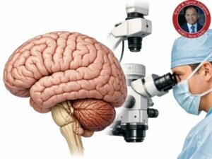
One of the most complicated and delicate operations in current medical practice is brain surgery. Every millimetre counts, and even the tiniest mistake can affect vital processes like memory, speech, or movement.
How, therefore, do Neurosurgeons get ready for such accuracy?
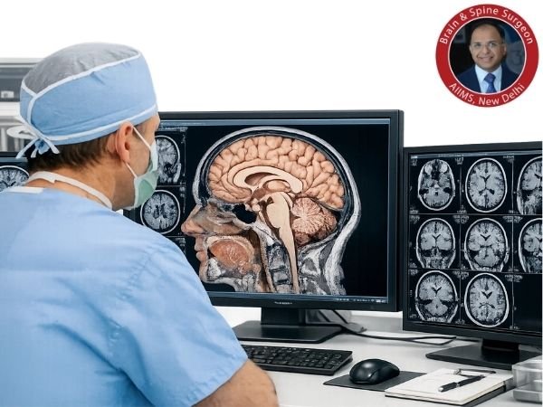
Doctors use sophisticated imaging, simulation tools, and specialised neurosurgery labs to thoroughly study the brain before surgery for hours or even days before entering the operation room. Better patient results and safer surgeries are guaranteed by this careful planning.
What is meant by “Studying the Brain Before Surgery”?
A combination of precise imaging, anatomical mapping, and surgical planning methods known as “pre-surgical brain study” enables neurosurgeons to:
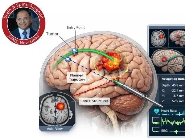
- Determine the precise site of the issue (tumour, AVM, aneurysm, etc.)
- Recognise how it relates to important brain regions.
- Choose the safest course for surgery.
- Forecast and reduce hazards
In neurosurgery, this procedure is frequently referred to as preoperative planning.
What Makes a Thorough Brain Study Crucial Before Surgery?
Everything is controlled by the brain, including personality and breathing. It is unable to handle even small harm, in contrast to other organs.
Among the main causes are:
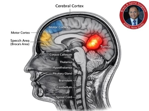
- Keep away of important areas including the speech and motor regions
- Lowering the likelihood of surgical complications including bleeding or neurological impairments
- Increasing accuracy when removing a tumour or lesion
- Improving the results of recovery
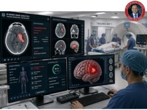
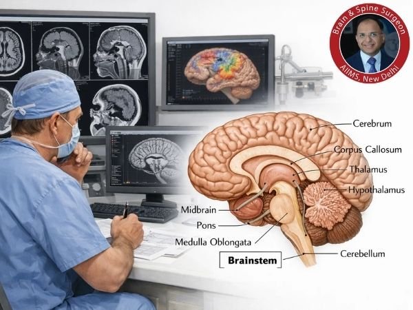
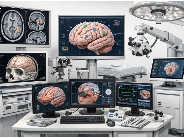
Technology and Equipment Used in Neurosurgery Laboratories
Modern neurosurgery labs have been equipped with modern equipment that enables medical professionals to see and model the brain in remarkable detail.
1. Magnetic resonance imaging, or MRI
High-resolution pictures of the soft tissues of the brain are produced by MRI.
Applications:
- Find abnormalities, infections, or tumours
- Determine any edema or structural alterations
2. fMRI, or functional MRI
fMRI displays brain activity in real time, in contrast to traditional MRI.
Why it’s important:
- Maps the regions involved in memory, movement, and speech
- Aids surgeons in preventing harm to these areas
3. Computed Tomography (CT) Scan
Bone and blood arteries can be seen in great detail using CT scans.
Utilised for:
- Finding fractures or bleeding
- Planning for emergency surgery
4. DTI, or diffusion tensor imaging
White matter tracts, or the brain’s communication channels, are mapped by DTI.
The significance:
- Maintains neuronal connections
- Crucial for tumour operations close to vital routes
5. Surgical navigation and 3D brain mapping
Using imaging data, physicians build 3D models of the patient’s brain.
Advantages:
- See the precise location of any anomalies
- Determine the safest course for surgery
- Boost the precision of surgery
6. Simulation and Virtual Reality (VR) Labs
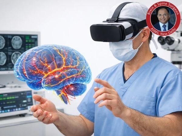
Surgeons can practise procedures in modern neurosurgery labs.
Benefits:
- Practice challenging situations
- Cut down on intraoperative surprises
- Boost self-assurance and accuracy
7. Labs for Cadaveric Brain Dissection
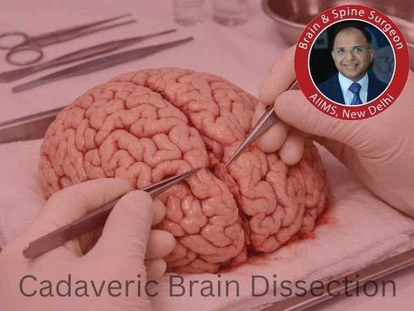
To have a thorough understanding of the structure of the brain, neurosurgeons frequently receive training in anatomical dissection labs.
Why it matters:
- Gives practical anatomical knowledge
- Improves knowledge of the spatial organization of brain structures
- Enhances surgical abilities
Related Post: How Neurosurgeons Learn Brain Anatomy: Role of Advanced Dissection Techniques
Step-by-Step: How Physicians Get Ready for Brain Surgery
- Step 1: Assessment of the patient
- Step 2: Imaging Research
- Step 3: Mapping the Brain
- Step 4: Planning for Surgery
- Step 5: Simulation (if required)
- Step 6: Discussion of the Final Strategy
Related Post: What to Expect Before and After Brain Surgery
Artificial Intelligence’s Place in Brain Research
AI is changing neurosurgery in the following ways:
- Analysing brain scans automatically
- Estimating the risks of surgery
- Supporting the identification and classification of tumours
- Improving surgical navigation accuracy
AI integration guarantees quicker and more precise decision-making.
How Patients Gain from This
These cutting-edge preparations directly assist patients:
- Safer procedures
- Decreased issues
- Improved brain function preservation
- Quicker recuperation times
Common Disorders Needing In-depth Brain Research
- Brain cancers
- AVMs, or arteriovenous malformations
- Aneurysms
- Epilepsy
- Brain damage caused by trauma
- Lesions at the base of the skull
Conclusion
Neurosurgery starts much earlier, in sophisticated neurosurgery labs, and is not limited to what takes place in the operating room.
Doctors make sure that every surgery is as safe and accurate as possible by using surgical simulations, advanced imaging, and in-depth brain research.
Neurosurgeons like Dr. Deepak Agrawal are able to provide exceptional, patient-centered care with the best results because of their careful preparation.
Sources:
- https://pmc.ncbi.nlm.nih.gov/articles/PMC3357627/
- https://pmc.ncbi.nlm.nih.gov/articles/PMC10030379/
- https://pmc.ncbi.nlm.nih.gov/articles/PMC3064825/
- https://pubmed.ncbi.nlm.nih.gov/12442617/
- https://pubmed.ncbi.nlm.nih.gov/37147485/
- https://pubmed.ncbi.nlm.nih.gov/27447756/
- https://pmc.ncbi.nlm.nih.gov/articles/PMC4523152/