Role of Transcranial Ultrasound and CT Scan in Detecting Brain Disorders
In order to avoid consequences including stroke, brain haemorrhage, tumours, and neurodegenerative illnesses, early detection of brain abnormalities is essential. Doctors can rapidly and precisely detect brain abnormalities using modern neuroimaging techniques.
Computed Tomography (CT) scans and transcranial ultrasound (also known as transcranial Doppler) are two frequently utilised diagnostic techniques. These technologies offer important insights into the anatomy, blood flow, and possible harm of the brain.
This page describes the function of CT scans and transcranial ultrasound brain diagnosis, as well as their advantages and recommended times.
Knowing Brain Disorders
Conditions that impact the structure, blood vessels, nerves, or function of the brain are referred to as brain disorders. These might appear gradually (like Parkinson’s disease) or abruptly (like a stroke).
Typical Brain Conditions
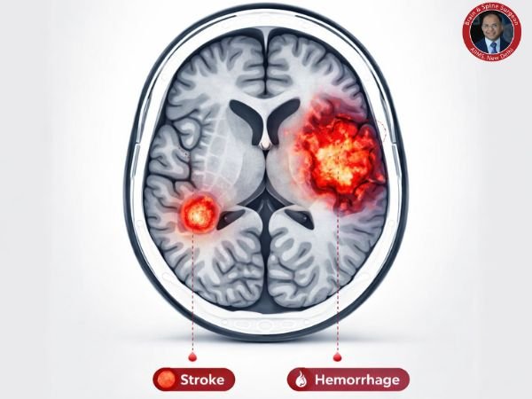
A stroke
Bleeding in the brain
Brain cancers
Hydrocephalus
Brain damage caused by trauma
Cerebrovascular injury
Neurodegenerative conditions
Early diagnosis of brain illnesses enables medical professionals to begin therapy promptly and enhance patient outcomes.
What is Transcranial Doppler, or Transcranial Ultrasound?
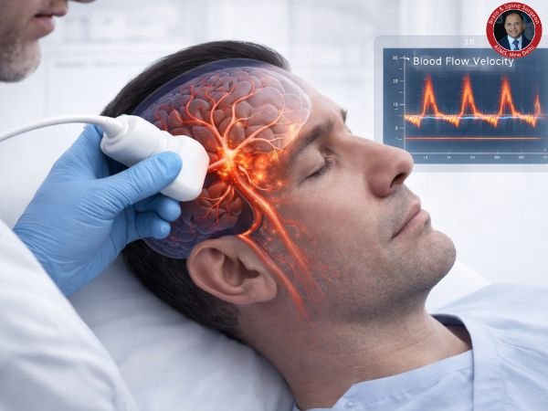
Transcranial ultrasound, often known as transcranial Doppler (TCD), is a non-invasive imaging diagnostic that assesses blood flow within the brain using sound waves.
A tiny probe passes ultrasonic waves through the skull throughout the examination. The speed and direction of blood flow in brain arteries can be measured by physicians thanks to these waves’ reflection off moving blood cells.
Transcranial ultrasonography, in contrast to CT or MRI examinations, primarily evaluates brain blood vessel circulation rather than brain tissue structure.
How Brain Disorders Can Be Found Using Transcranial Ultrasound
1. Identifying the Risk of Stroke
One of the main causes of stroke is constricted or obstructed brain arteries, which can be found with transcranial ultrasonography.
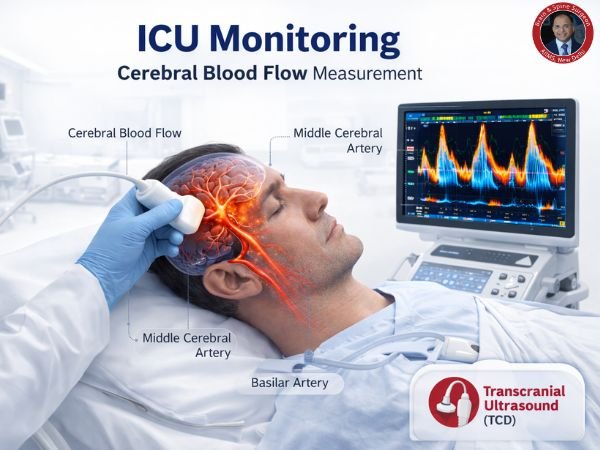
2. Tracking the Brain’s Blood Flow
It assists medical professionals in identifying circulatory irregularities by measuring the blood flow velocity in arteries like the Circle of Willis.
3. Identifying Vasospasm
Vasospasm is the abrupt narrowing of arteries following disorders such as subarachnoid haemorrhage. This issue is monitored with the aid of transcranial Doppler.
4. Embolism Detection
It is possible to identify tiny clots that are moving toward the brain from the heart or neck veins.
5. Keeping an Eye on Brain Conditions
It can assist in keeping an eye on:
Brain aneurysm
TIA (transient ischaemic attack)
Variations in intracranial pressure
Risk of stroke from sickle cell disease
What is a Brain CT Scan?
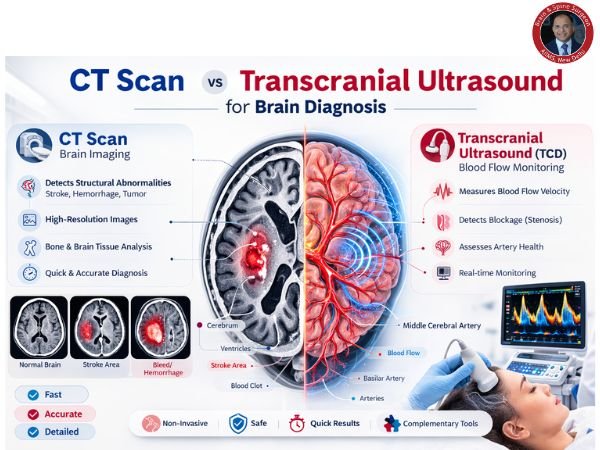
A Computed Tomography (CT) scan is a diagnostic imaging method that produces finely detailed cross-sectional images of the brain using X-rays and computer processing.
Clear information regarding brain architecture, haemorrhage, swelling, tumours, and traumas can be obtained from CT scans.
Because of their rapid outcomes, they are frequently employed in neurological emergencies.
CT Scan’s Function in Identifying Brain Disorders
1. Recognising Brain Haemorrhage
The first-line test for identifying cerebral haemorrhage, particularly following a head injury, is a CT scan.
2. Brain Tumour Detection
Know More: Life After Brain Tumour Surgery: Recovery, Care and Lifestyle Changes
They aid in locating and identifying abnormal growths.
3. Traumatic Brain Injury Diagnosis
CT imaging rapidly identifies:
Fractures of the skull
Swelling of the brain
Clots of blood
4. Assessing Stroke
CT scans assist in distinguishing between:
Ischaemic stroke (artery blockage)
Hemorrhagic stroke (brain haemorrhage)
5. Identifying Hydrocephalus
It aids in determining whether the brain has accumulated too much cerebrospinal fluid.
CT Scan vs. Transcranial Ultrasound
| Feature | Ultrasound of the Brain | Standard CT Scan |
|---|---|---|
| Technological advances | Waves of sound | X-ray pictures |
| What is assessed | Brain artery blood flow | Structure of the brain |
| Radiation | Absence of radiation | Makes use of radiation |
| Velocity/Speed | Quick test at the bedside | Quick imaging in hospitals |
| Utilise | Keeping an eye on circulation | Identifying structural problems |
Because these tests are complimentary, a precise diagnosis is frequently achieved by combining them.
Transcranial Ultrasound Benefits
Painless and non-invasive
Absence of radiation exposure
Bedside-friendly and portable
Permits ongoing observation
Fast and economical
Because transcranial sonography may be performed regularly to evaluate brain abnormalities, it is very helpful in intensive care units.
When Are These Tests Recommended by Doctors?
A neurosurgeon or neurologist may suggest transcranial ultrasonography or CT scan if a patient has:
Abrupt, intense headache
Diminished awareness
Symptoms of a stroke
Head trauma
Numbness or weakness in the limbs
Speech or vision issues
Possible brain tumour or haemorrhage
These tests aid in verifying the diagnosis and directing the course of treatment.
How the Examinations Are Conducted
The Process of Transcranial Ultrasound
The neck or temples are treated with a gel.
Sound waves are sent via a tiny probe (transducer).
The device logs patterns of blood flow.
Typically, the test takes 30 to 60 minutes.
The Process of a CT Scan
On a moving table, the patient lies.
The head is rotated by the CT scanner.
Several pictures are taken.
Typically, the procedure takes 5-10 minutes.
Transcranial Ultrasound’s Drawbacks
Transcranial ultrasonography is helpful, but it has drawbacks.
Image quality may be limited by the thickness of the skull
Unable to display intricate brain structures
Frequently utilised when used with CT or MRI
For a thorough assessment, physicians frequently combine Transcranial Ultrasound Brain Diagnosis with CT or MRI.
The Prospects for Brain Imaging
Improvements in medical imaging are making it possible to identify brain problems earlier and more precisely.
Among the emerging technologies are:
AI-powered brain imaging
Ultrasound with high resolution
Devices for portable neuroimaging
These developments could greatly enhance neurological disease early diagnosis and therapy.
When Is the Right Time to Consult a Neurosurgeon?
Get medical attention right away if you encounter:
Abrupt, intense headache
One-half of the body is weak
Vision loss
Speaking difficulties
Persistent lightheadedness
Confusion due to a head injury
Early diagnosis of brain illnesses can avert irreversible brain damage and save lives.
In Conclusion
In order to avoid major neurological problems, early detection of brain abnormalities is essential.
While CT scans offer precise structural images of brain tissues, transcranial ultrasound brain diagnosis aids medical professionals in assessing blood circulation in the brain.
When combined, these tools improve patient outcomes by enabling neurosurgeons to promptly diagnose brain diseases and initiate appropriate treatment.
FAQ’s
1. What is transcranial ultrasonography used for?
By assessing blood flow in the brain’s arteries, transcranial ultrasonography can identify circulatory irregularities, vasospasm, and stroke risk.
2. Is it safe to use transcranial ultrasound?
Indeed. It is a painless and safe test since it use sound waves rather than radiation.
3. What conditions of the brain may CT scans identify?
Brain bleeding, tumours, stroke, skull fractures, hydrocephalus, and traumatic brain injuries can all be detected by CT scans.
4. For brain diagnosis, which is preferable: an ultrasound or a CT scan?
They have distinct functions. While transcranial ultrasonography assesses blood flow, CT images display the structure of the brain.
5. What is the duration of a CT scan of the brain?
A CT scan is helpful in emergency situations because it typically takes five to ten minutes.
6. Can tumours be seen with transcranial ultrasound?
Although it is mostly used for blood flow measurement rather than precise tumour detection, it may identify some anomalies.
Sources: