A Choroidal Melanoma Tumour: What Is It?
An uncommon kind of eye cancer called a choroidal melanoma tumour arises in the choroid, a layer of blood vessels and connective tissue situated between the retina and the sclera (the white portion of the eye).
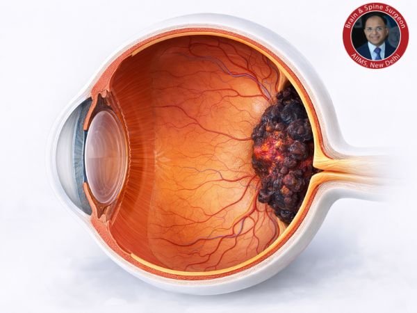
Melanocytes, which produce pigment, are the source of this type of uveal melanoma, which is the most prevalent primary intraocular (within the eye) malignancy in people.
Despite being uncommon, it is a dangerous disorder that needs prompt diagnosis and treatment in order to protect vision and stop its progression.
Where Is Choroidal Melanoma Found?
The uvea has three layers of the eye:
- Iris (coloured portion)
- The ciliary body
- Choroid (most frequently impacted)
The choroid is an essential structure for vision because it provides the retina with oxygen and nutrients.
Risk Factors and Causes
Although the precise aetiology of choroidal melanoma is not always known, a number of risk factors raise the risk:
- Age (more prevalent among middle-aged and older persons)
- Light-colored eyes and fair skin
- Overexposure to UV light
- Mutations in genes
- Ocular nevi, or freckles, are present
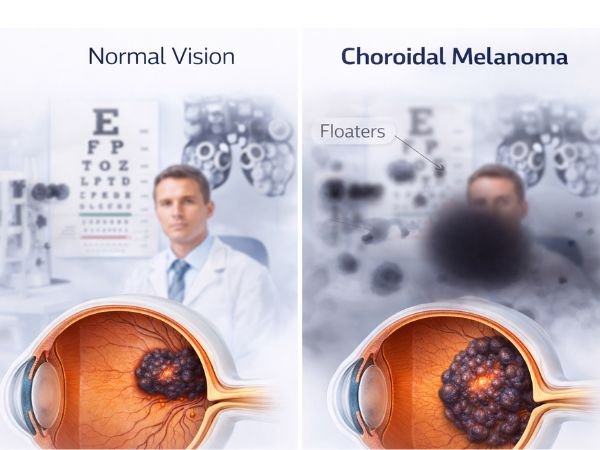
Choroidal Melanoma Symptoms
Many people may not show any symptoms in the early stages. The following symptoms could appear as the cancer grows:
- Vision deformation or blurriness
- Peripheral vision loss
- Light flashes (photopsia)
- Floaters (black areas in the field of vision)
- Rarely, a black patch on the iris is visible
- Pain (in more severe situations)
How is the Diagnosis of Choroidal Melanoma Made?
Early detection is essential. Typically, a diagnosis includes:
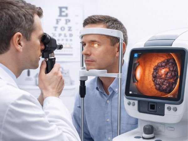
1. Eye Exam
A thorough examination of the eyes using ophthalmoscopy to see the tumour.
2. Imaging Examinations
- B-scan ultrasound
- OCT (optical coherence tomography)
- Fundus photography
3. High-Tech Imaging
- CT or MRI scan (to evaluate spread)
4. Biopsy (in rare instances)
Occasionally carried out to evaluate genetic risk or confirm a diagnosis.
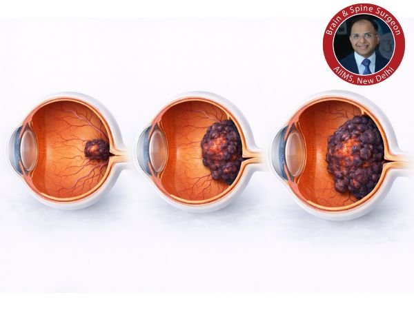
Is Choroidal Melanoma Hazardous?
Indeed, if left untreated, choroidal melanoma can be fatal.
- It might develop and impair vision
- It has the potential to metastasise, particularly to the liver
- Early intervention greatly enhances results
Options for Choroidal Melanoma Treatment
The size, location, and general health of the patient all influence the course of treatment.
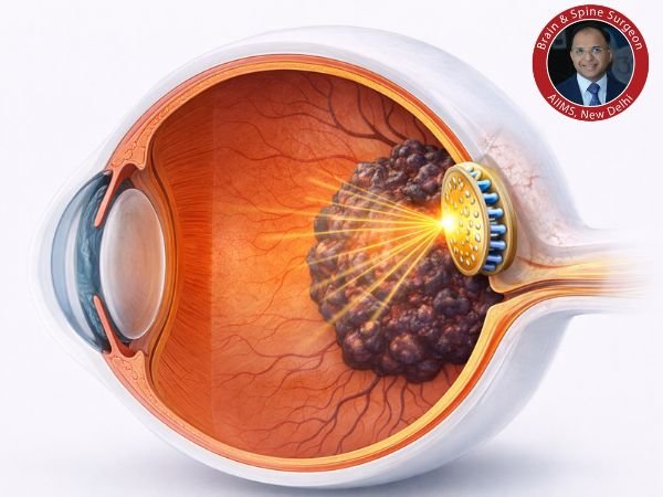
1. Most Common: Radiation Therapy
Brachytherapy for Plaque
- A tiny radioactive disc is positioned close to the tumour
- Delivers specific radiation
Treatment Using Proton Beams
- Extremely accurate radiation
- Reduces harm to nearby tissues
2. Laser Treatment
Thermotherapy Transpupillary (TTT)
- Uses an infrared laser to reduce the size of tiny tumours
3. Local Resection Surgery
- Tumour removal with keeping the eye intact
Enucleation
- Removal of the entire eye (in cases of big tumours)
- When vision cannot be preserved
4. Systemic Therapy
If the cancer spreads:
- Immunotherapy
- Targeted treatment
- Chemotherapy (less successful for melanoma)
Recuperation Following Therapy
The type of treatment determines recovery:
- Radiation therapy requires frequent follow-ups and gradual progress
- Surgery: After enucleation, prosthetic eyes may be used; healing times vary
- Vision outcome: May be partially or fully affected
To find recurrence or metastasis, routine monitoring is essential.
Issues to Be Aware of
- Loss of vision
- Detachment of the retina
- Retinopathy caused by radiation
- Recurrence of the tumour
- Metastasis, particularly with regard to the liver
Is It Possible to Prevent Choroidal Melanoma?
Although prevention is impossible, risk can be decreased by:
- Putting on UV-blocking sunglasses
- Frequent eye exams
- Keeping a watch on eye moles (nevi)
- Early medical attention for changes in vision
Survival and Prognosis
The prognosis is based on:
- Size of the tumour
- Profile of genetics
- Early identification
When small tumours are discovered early, their survival rate is significantly higher than that of advanced instances.
When Is It Time to See a Physician?
Speak with an expert right away if you encounter:
- Abrupt shifts in vision
- Flashes or persistent floaters
- Dark areas in the field of vision
- Pain or discomfort in the eyes
Both life and vision can be saved by an early diagnosis.
In conclusion
When detected early, a choroidal melanoma tumour is a dangerous but treatable eye cancer. Symptom awareness, prompt diagnosis, and suitable therapy can greatly enhance results and protect vision.
Getting professional medical help as soon as you notice any strange changes in your vision can make all the difference.
FAQ’s
1. Is it possible to treat choroidal melanoma cancer?
Yes, it can be successfully treated if found early. Advanced instances, however, have the potential to spread and prove fatal.
2. How common is choroidal melanoma?
No, it is the most prevalent intraocular cancer in adults, although it is an uncommon type of eye cancer.
3. Is it possible for it to spread to other body parts?
Indeed, particularly to the liver. Frequent follow-ups are crucial.
4. Will I get blind?
Not all the time. While advanced cases may result in vision loss, early treatment may save vision.
5. Is surgery always necessary?
No, radiation therapy is used to treat many situations without removing the eye.
6. How frequently should a follow-up be conducted?
Usually every three to six months at first, then once a year based on risk.
Sources: