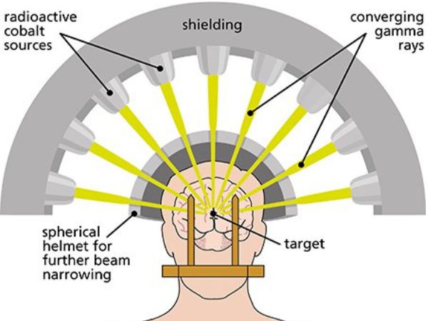
Treatment for a variety of brain illnesses, including vascular malformations, brain tumours, and functional problems like trigeminal neuralgia, has been transformed by Gamma Knife Radiosurgery (GKRS). GKRS, which is well-known for its accuracy and non-invasiveness, targets and eliminates abnormal tissue while protecting nearby healthy brain structures with concentrated radiation beams.
It does, however, include some risk, just like any medical operation. Understanding the potential Adverse Radiation Effects (AREs) after treatment is one of the most crucial factors for both patients and clinicians.
The definition, causes, symptoms, diagnosis, treatment, and ways to reduce AREs are all covered in this article.
Adverse Radiation Effects (AREs): What Are They?
Unwanted changes in brain tissue brought on by the high-dose, concentrated radiation used in Gamma Knife Radiosurgery are known as Adverse Radiation Effects (AREs). Even though GKRS reduces exposure to nearby tissues, radiation harm might cause short-term or long-term negative effects in certain people.
From minor swelling and temporary neurological symptoms to more severe side effects like radionecrosis (radiation-induced tissue death), these consequences can vary widely.
Adverse Radiation Effect Types
Acute Impacts (Days to Weeks)
- Rare with Gamma Knife
- May consist of localised swelling, headache, or nausea.
- Caused by transient swelling or inflammation at the treatment location
- Subacute Side Effects: 1–6 Months Following Therapy
Common
- Incorporate fatigue, slight cognitive impairments, or localised brain swelling.
- Typically reversible and conservatively handled
- Late Effects: Six Months to Years Following Therapy
May consist of:
- Dead brain tissue brought on by radiation damage is known as radiation necrosis.
- Swelling: a buildup of fluid that results in symptoms and pressure
- Calcification or cyst development in the treated area
- Memory issues, seizures, or delayed neurological deficiencies
What Leads to AREs?
The probability of getting AREs is affected by multiple factors, including:
- Radiation Dose: Risk is increased by higher dosages or greater treatment volumes.
- Lesion location: Eloquent brain regions, optic pathways, and the brainstem are particularly sensitive.
- Lesion Size: Greater treatment volumes are needed for larger lesions.
- Tissue sensitivity is increased by prior radiation exposure.
- Patient-specific factors include genetic vulnerability, age, and medical comorbidities.
Adverse Radiation Effect Symptoms
Depending on the part of the brain that is impacted, symptoms can include:
- A headache
- Vomiting or feeling queasy
- Limb weakness or numbness
- Seizures
- Having trouble understanding or speaking the language
- Changes in vision
- Cognitive decline or memory loss
Recognising Adverse Effects of Radiation
To distinguish AREs from tumour recurrence or disease progression, a prompt and precise diagnosis is crucial. Among the tools utilised are:
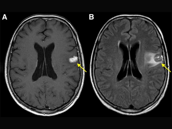
- MRI with contrast: Displays changes and swelling in the treated area
- MR Spectroscopy or Perfusion Imaging: Assists in distinguishing between tumour recurrence and radiation necrosis
- PET scans are occasionally used to evaluate brain metabolism.
- Clinical Evaluation: History of symptoms and neurological examination
Control and Handling of AREs
The intensity of the symptoms determines the course of treatment:
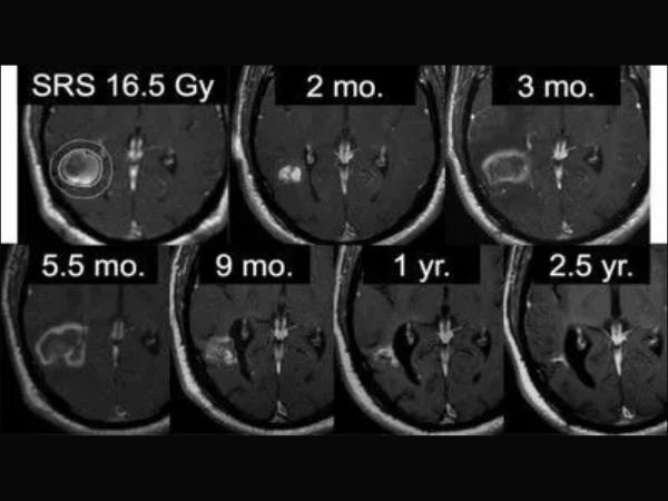
- Observation
- Numerous AREs self-limit and are tracked by routine MRIs.
- Drugs
- Steroids help lessen brain oedema and inflammation, such as dexamethasone
- If seizures happen, anti-seizure drugs
- Supportive care and pain management
- Bevacizumab
- A targeted treatment that inhibits VEGF, a protein that causes new blood vessel growth and edema, decreases swelling and improves radiation necrosis symptoms
- Surgery
- Although rarely required, it could be carried out in cases of severe necrosis, cyst formation, or unclear diagnosis.
How to Reduce the Negative Effects of Radiation
- Selecting the appropriate GKRS candidates with consideration for tumour size, location, and general health
- Improved Treatment Planning: Sophisticated software and imaging technologies for precise dosage management
- In certain situations, fractionated radiosurgery may involve administering radiation in lesser doses over a number of sessions.
- Close Monitoring: Following treatment, routine MRIs and clinical evaluations
In conclusion
Gamma Knife particularly for patients with deep-seated or physically inaccessible brain lesions, radiosurgery is still a safe and useful option in the neurosurgical toolbox. However, for well-informed decision-making, it is essential to understand the possibility of adverse radiation effects.
The majority of AREs can be successfully controlled with careful planning, consistent observation, and prompt action, enabling patients to confidently take advantage of the accuracy and strength of Gamma Knife Radiosurgery.
Sources:
- https://www.ahajournals.org/doi/10.1161/STROKEAHA.116.014825#sec-3
- https://pubmed.ncbi.nlm.nih.gov/37026335/
- https://journals.lww.com/neur/fulltext/2023/71001/adverse_radiation_effects_following_gamma_knife.10.aspx
- https://my.clevelandclinic.org/health/procedures/16559-gamma-knife-surgery
- https://pubmed.ncbi.nlm.nih.gov/27903185/