Skip to content

Gamma Knife Radiosurgery for Atypical Meningioma

Topics

Table of Contents

Overview

Atypical Meningioma
Atypical Meningioma

One of the most typical kinds of brain tumors is a meningioma, which develops from the meninges, which are the protective coverings that surround the brain and spinal cord. Some meningiomas can be more aggressive, although the majority are benign and grow slowly. Atypical meningioma, which is categorised as WHO Grade II, is one such variation. Compared to normal (benign) meningiomas, these tumours have a higher chance of recurrence and need to be managed more carefully.

Atypical Meningioma: What Is It?

A brain tumour that develops from the meninges but exhibits characteristics that make it more aggressive than a conventional (Grade I) meningioma is known as an atypical meningioma. These cancers:

Atypical Meningioma (2)
  • Possess a greater cell division rate.
  • Can infiltrate adjacent brain tissue
  • They are more likely to return following therapy.
  • Represent roughly 15–20% of all meningiomas.

Reasons and Risks

Although the precise cause of atypical meningiomas is unknown, the following risk factors are linked to their development:

  • Exposure to radiation
  • Hereditary disorders such as neurofibromatosis type 2 (NF2)
  • Effects of hormones, particularly in women
  • Meningioma family history

Signs and Symptoms

Symptoms of atypical meningiomas depend on their size and location in the brain or spinal cord. Typical signs and symptoms include:

A headache

  • Seizures
  • Issues with vision
  • Deafness
  • Numbness or motor weakness
  • Changes in personality or cognition

Compared to normal meningiomas, these tumours have a faster rate of growth, which allows them to produce symptoms more quickly.

Diagnosis

Atypical meningioma diagnosis includes:

  1. A neurological examination evaluates the function of the brain and nerves.
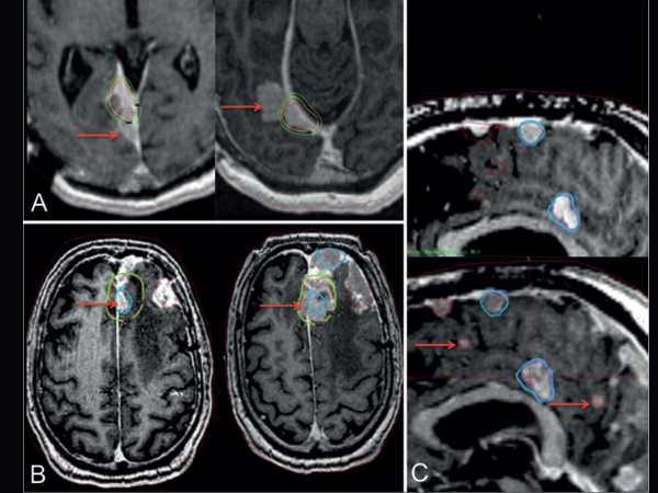
  1. Imaging Research:
  • The preferred method for visualising tumours is magnetic resonance imaging, or MRI.
  • To find any bone involvement, use a CT scan.
  1. Surgical resection or biopsy: for a clear diagnosis based on histological analysis.

Under a microscope, characteristics like enhanced mitotic activity, necrosis, and brain invasion aid in the tumor’s classification as atypical.

Options for Treatment

  1. Surgery

Usually, the first course of treatment is surgical removal. Gross total resection (GTR), or the total excision of the tumour, is the aim. However, total removal might not always be feasible because of the tumor’s aggressive nature or close proximity to important brain regions.

  1. Treatment with Radiation

After surgery, radiation is frequently utilised, particularly if:

  • The tumour was not entirely eliminated.
  • The tumour exhibits aggressive traits.
  • Recurrence is present.

This includes more accurate choices like stereotactic radiosurgery as well as traditional external beam radiation therapy.

Radiosurgery with Gamma Knife for Atypical Meningioma

Radiosurgery with Gamma Knife for Atypical Meningioma

A non-invasive, highly targeted type of radiation therapy, Gamma Knife Radiosurgery (GKRS) is particularly useful in treating atypical meningiomas, specifically:

  • Tumours that are found in sensitive or deep brain areas
  • Tumours that remain after surgery
  • Periodic meningiomas
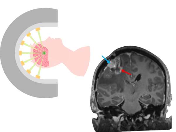
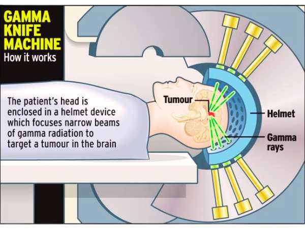
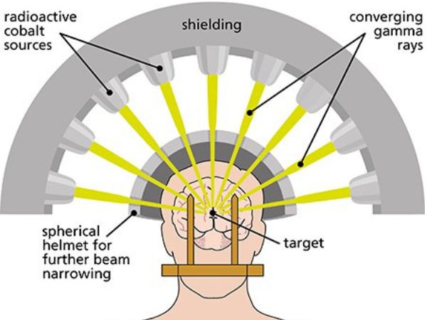
How It Operates: 

  • Targets the tumour with several gamma radiation beams.
  • Minimally harms the healthy tissue around it.
  • Usually finished without requiring a surgical incision in a single session.

Gamma Knife Radiosurgery Advantages:

Gamma Knife Radiosurgery
  • Non-invasive: neither general anaesthesia nor open surgery are required.
  • Patients usually return home the same day after an outpatient operation.
  • Accurate targeting lessens harm to important brain regions.
  • Reduced rate of complications in contrast to traditional surgery.
  • A beneficial supplement to surgery or a substitute in cases where surgery is not feasible.

Limits:

  • Not appropriate for extremely big tumours.
  • For tumours that are spreading quickly or are highly invasive, surgery might not be the best option.
  • To determine whether there has been a recurrence or progression, long-term monitoring is necessary.

Prognosis and Follow-up

Even after total surgical removal or radiosurgery, patients with atypical meningiomas need to have regular MRI scans performed as part of a long-term follow-up. At 20% to 40% within 5 years, the recurrence rate is much higher than that of benign meningiomas.

The outcome is dependent on:

  • The degree of resection
  • Features of histology
  • Reaction to radiation treatment
  • The patient’s overall or general health

In conclusion

Because of their aggressive nature and tendency to recur, atypical meningiomas pose particular difficulties. The best possibilities for long-term control are with a mix of radiation and surgery, such as Gamma Knife Radiosurgery. It is essential to speak with a qualified neurosurgeon and a multidisciplinary team if you or a loved one has been diagnosed with an atypical meningioma in order to create a customised treatment strategy.

Please get in touch with our centre for professional treatment of atypical meningiomas, including the newest Gamma Knife technologies.

Sources:

  1. https://pmc.ncbi.nlm.nih.gov/articles/PMC7714950/
  2. https://pubmed.ncbi.nlm.nih.gov/32586495/
  3. https://radiopaedia.org/articles/atypical-meningioma
  4. https://www.pathologyoutlines.com/topic/cnstumoratypicalmeningioma.html
  5. https://www.sciencedirect.com/science/article/abs/pii/B9780128221983000434
  6. https://www.advancesradonc.org/article/S2452-1094(24)00272-0/fulltext
  7. https://www.sciencedirect.com/science/article/abs/pii/S1878875015013649
  8. https://link.springer.com/chapter/10.1007/978-3-7091-1376-9_13
  9. https://www.sciencedirect.com/science/article/pii/S2452109424002720
  10. https://www.redjournal.org/article/S0360-3016(09)01590-9/fulltext
Table of Contents

You May Also Know Related to Gamma Knife
FAQ’s

Gamma Knife uses 192–201 precisely focused beams of gamma rays that converge at a single target point in the brain. This high dose of radiation damages the DNA of abnormal cells, stopping their growth or causing them to shrink, while minimizing exposure to nearby healthy tissue.

No, Gamma Knife is not a traditional surgery. It is a non-invasive outpatient procedure performed without any incision, stitches, or general anesthesia in most cases.

Gamma Knife is commonly used for:

Brain tumors (benign and malignant)
Metastatic brain lesions
Arteriovenous malformations (AVMs)
Trigeminal neuralgia
Acoustic neuromas (vestibular schwannomas)
Pituitary tumors
Certain functional disorders (like tremors)

The procedure is generally painless. Patients may feel mild pressure while the head frame (if used) is applied or may experience slight discomfort from anesthesia injections, but the treatment itself is not painful.

Depending on the condition and treatment plan, Gamma Knife may take 30 minutes to 3 hours. Most patients go home the same day.

Common side effects are usually mild and temporary, including:

Headache
Nausea
Fatigue
Mild scalp swelling or tenderness at the frame site
Rarely, some patients may experience delayed radiation effects such as brain swelling or radiation necrosis.

Most patients can return to their normal routine within 24–48 hours after the procedure.
You may feel mild fatigue for a few days.
Follow-up imaging is usually required after 1 year.

Gamma Knife has a high success rate and has been used worldwide for decades. Its effectiveness depends on the condition treated, size and location of the lesion, and overall patient health. In many cases, it offers results comparable to open surgery with fewer risks.

Yes, in some cases Gamma Knife treatment can be repeated if the disease recurs or if new lesions develop.

Yes. Gamma Knife is considered one of the safest and most precise forms of radiosurgery, with millions of patients treated globally and extensive clinical data supporting its use.

Eligibility depends on factors such as:

Size and location of the lesion
Overall health and age
Whether open surgery is too risky
A neurosurgeon and radiation oncologist will decide if Gamma Knife is the best option for you.

A detailed MRI or CT scan is performed to map the brain.
A lightweight head frame or mask is used for accuracy.
Your doctors plan the radiation dose and target areas using specialized software.

Local anesthesia is given if a head frame is used.
General anesthesia is usually not required, except for children or patients unable to remain still.

Costs vary depending on hospital, city, and condition treated. On average, Gamma Knife in India ranges from ₹1.5 lakh to ₹4.5 lakh. It is usually more affordable compared to treatment in Western countries. At AIIMS Delhi, it is much more affordable around ₹75,000 which makes it affordable for the patients in need.

AIIMS Delhi is one of the leading centres for Gamma Knife in India.

AIIMS uses the latest Gamma Knife Perfexion system.

It has successfully treated thousands of patients for brain tumors, blood vessel problems, and even eye cancers.

The treatment is done by expert neurosurgeons such as Dr. Deepak Agrawal and team.

Clinic timings for Gamma Knife OPD at AIIMS Delhi: Monday & Friday, 8:00 AM – 9:00 AM.

Cost is around ₹75,000 and is subsidized compared to private hospitals.

The whole procedure is usually done in one day:

  • Consultation & Planning – Doctors review your scans and decide the treatment.
  • Head Frame/Mask – A lightweight frame or mask is placed to keep your head still.
  • Imaging – MRI/CT scans are done to locate the exact target.
  • Treatment Planning – Specialists plan the dose and direction of radiation.
  • Gamma Knife Treatment – You lie on the machine; painless radiation beams treat the target (30 mins–3 hrs).
  • Recovery & Discharge – The frame is removed, and most patients go home the same day.
  • Follow-up – MRI after a few months to check results.

Before you get a treatment date, you need to complete a few steps:

  • OPD Registration – Visit the Neurosurgery OPD (Gamma Knife clinic at AIIMS, Mon & Fri 8–9 AM) and register.
  • Consultation with Doctor – Meet the neurosurgeon who will check your reports, MRI/CT scans, and medical history.
  • Investigations – Sometimes fresh MRI/CT or blood tests are needed for treatment planning.
  • Medical Fitness – Basic checks like blood pressure, sugar, heart condition, etc.

Financial/Insurance Approval –

  1. If using Ayushman Bharat or government schemes, you need approval papers.
  2. If self-paying, you will be given the estimated cost (~₹75,000 at AIIMS).
  • Consent & Admission Slip – Once doctors confirm you are fit and formalities are done, you sign the consent form.
  • Treatment Date Allotment – A date is given for your Gamma Knife procedure.

Once you receive your Gamma Knife date, you’ll need to do a few simple things before the procedure:

  • Admission Process – Report to AIIMS on the morning of your procedure and complete admission at the Neurosurgery ward/Daycare.
  • Paperwork – Carry your OPD slip, MRI/CT films, blood test reports, admission slip, and ID proof. If you are covered under Ayushman Bharat/insurance, keep those approval papers ready.
  • Consent Forms – You (or a family member) will sign consent for the procedure and anesthesia.
  • Medical Check-up – Doctors will check your BP, sugar, heart rate, and do a quick physical exam.
  • Fasting – Usually, you will be asked to not eat or drink for 6 hours before the procedure.
  • Pre-Procedure Prep – An IV line may be put, and medicines (sedation/antibiotics if needed) are given.
  • Treatment – You are then taken to the Gamma Knife room for frame/mask placement, imaging, and treatment.

After the procedure, you’ll be observed for a few hours and most patients are discharged the same day or next morning.

No, You can have light breakfast.

Yes, you can take the medicine and then come for the treatment.