Recognising a potential development in artificial intelligence and ocular ultrasound-based brain monitoring
In patients with severe brain disorders such as traumatic brain injury (TBI), brain swelling, intracranial haemorrhage, hydrocephalus, and other causes of elevated intracranial pressure, intracranial pressure (ICP) is a crucial measure. Significant increases in ICP can impair blood flow to the brain and, in extreme situations, result in irreversible brain damage or even be fatal.
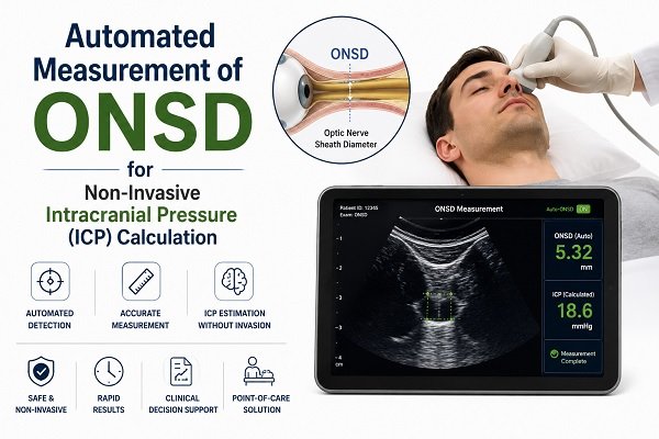
Traditionally, an invasive technique involving the implantation of a pressure-monitoring device inside the skull has been necessary for accurate continuous ICP monitoring. These procedures are invasive and call for specialised knowledge, even though they are still the clinical reference standard for suitable patients.
Optic nerve sheath diameter (ONSD) assessment with ocular ultrasonography is a possible substitute. ONSD can now be automatically measured from ultrasound images, thanks to recent developments in image processing, automation, and artificial intelligence. This could make non-invasive evaluation of elevated ICP quicker, more reliable, and simpler.
In “A Novel Method for Prediction of Raised Intracranial Pressure Through Automated ONSD and ETD Ratio Measurement From Ocular Ultrasound,” Dr. Deepak Agrawal of AIIMS, New Delhi, investigated an automated method that combines ONSD with eyeball transverse diameter (ETD) to improve prediction of raised ICP.
Intracranial Pressure (ICP): What Is It?
The skull is a closed structure made up of three main parts:
- Brain tissue
- Blood
- CSF, or cerebrospinal fluid
Intracranial pressure (ICP) is the pressure that these materials cause inside the skull.
The body normally keeps ICP within a somewhat controlled range. On the other hand, diseases like hydrocephalus, brain enlargement, brain tumours, infections, severe head trauma, and internal bleeding can raise intracranial pressure.
A substantially raised ICP could result in:
- Decreased cerebral blood flow
- Decreased brain tissue oxygen supply
- Swelling of the brain
- Changes in awareness
- In severe circumstances, brain herniation
Thus, in neurocritical care, early detection of elevated intracranial pressure is crucial.
Why Is It Important to Measure ICP?
ICP monitoring aids medical professionals in comprehending how the brain reacts to damage and therapy.
Continuous pressure measurements can be obtained with invasive monitoring for certain severely ill patients. Nevertheless, implanting an intracranial pressure monitor necessitates an invasive process and is often carried out in specialised conditions.
This raises a crucial clinical query:
Is it possible to detect or screen for elevated ICP without inserting a device into the skull?
Ocular ultrasound-based ONSD measurement is one possible solution.
ONSD: What Is It?
Optic Nerve Sheath Diameter is referred to as ONSD.
The brain and eye are connected by the optic nerve. The optic nerve sheath, which is contiguous with the membranes encircling the brain, envelops it.
Crucially, there is communication between the cerebral subarachnoid space and the area around the optic nerve. The optic nerve sheath may enlarge as a result of pressure being conveyed along this channel when intracranial pressure increases.
This implies that variations in the optic nerve sheath’s diameter may serve as a proxy for variations in intracranial pressure.
ONSD ultrasonography, a non-invasive method of measuring intracranial pressure, is based on this anatomical relationship.
How Is ONSD Calculated?
Ocular or transorbital ultrasound can be used to assess ONSD.
Using the proper coupling medium, a qualified medical practitioner carefully inserts an ultrasonic probe above the closed eyelid. It is then possible to see the optic nerve and its sheath behind the eyeball.
A standard distance behind the globe is used to estimate the diameter of the optic nerve sheath. Although measurement methods and procedures differ between research, measurements taken approximately 3 mm behind the eyeball have historically been widely employed.
Standardised imaging methods are crucial since even little variations in probe position, imaging plane, measurement location, and image quality might impact the outcome. Both eyes may be evaluated.
In order to standardise ONSD point-of-care ultrasound acquisition and measurement, international experts have recently created consensus-based quality criteria.
What Does a Greater ONSD Signify?
Generally speaking, an enlarged ONSD may be linked to elevated intracranial pressure since it may imply greater pressure surrounding the optic nerve.
For patients and their families, there is one crucial point, though:
No single ONSD result can reliably identify elevated ICP in all patients.
Different thresholds have been reported by various research. The measurement method, patient population, equipment, operator experience, and clinical state are some of the factors that can affect the value.
Although research has revealed thresholds ranging from below 5 mm to above 6 mm, a generally discussed barrier has typically been around 5 mm. According to a more recent meta-analysis, in certain situations, readings between 5.6 and 6.3 mm may offer more specificity. Therefore, it is not appropriate to use these figures as universal diagnostic cut-offs.
Why Measure ONSD Automatically?
A skilled operator must locate the optic nerve sheath on an ultrasound image and manually position measurement markers in order to do a traditional ONSD test. Operator-dependent variation may result from this.
By enabling computer algorithms to recognise important anatomical features and compute measures reliably, automated measurement seeks to lessen this variability.
An automated system could possibly:
- Get a video or picture of an ocular ultrasound.
- Determine the eyeball.
- Determine the location of the optic nerve and its sheath.
- Find the pertinent measurement points automatically.
- Determine ONSD.
- Calculate the transverse diameter (ETD) of the eyeball.
- Determine the ratio of ONSD to ETD.
- Indicate whether the measurements point to a greater chance of elevated ICP.
This method integrates clinical neurology, artificial intelligence/machine learning, image processing, and ultrasound imaging.
What Does the ONSD-to-ETD Ratio Mean?
The fact that ONSD might not be enough to predict elevated ICP is one of the significant advances investigated in the study involving Dr. Deepak Agrawal.
Thus, the study examined the relationship between Optic Nerve Sheath Diameter (ONSD) and Eyeball Transverse Diameter (ETD). The ratio is known as the ONSD-to-ETD ratio (OER).
To put it simply: OER = ONSD ÷ ETD
In comparison to ONSD alone, a ratio may assist adjust for individual variability in eye size and possibly give a more standardised biomarker.
What Were the Findings of Dr. Deepak Agrawal and Associates’ Research?
An automated methodology for calculating ONSD and ETD from ocular ultrasound images in trauma patients was assessed in the study, which was published in Ultrasonic Imaging.
In order to compare automated ultrasound-based assessments with traditionally measured ICP, the researchers examined data from 57 trauma patients at AIIMS, New Delhi.
According to the researchers:
- ONSD and ETD were measured using automated techniques.
- Compared to ONSD alone, the ONSD-to-ETD ratio (OER) had a higher connection with ICP.
- OER and ICP showed a correlation of r = 0.81 (p ≤ .01).
- ICP and ONSD alone had a correlation of r = 0.69 (p = .004).
- In this specific study cohort, an OER threshold of 0.21 was found to be predictive of elevated ICP.
- In the study dataset, patients with OER ≥0.21 had ICP readings between roughly 17 and 35 mmHg.
These results imply that automated ONSD and ETD measures could offer a helpful non-invasive method for identifying and keeping an eye on individuals who might have elevated ICP.
The study’s upper limit, however, shouldn’t be regarded as a clinical cut-off that applies to all patients. Before such automated biomarkers can be regarded as definitive substitutes for invasive ICP monitoring, larger and more varied investigations are required.
Why Could the ONSD-to-ETD Ratio Be Significant?
ONSD can differ from person to person. As an example, two individuals may naturally differ in the size or anatomical features of their eyeballs. Individual variance may consequently have an impact on a measurement based solely on ONSD. It could be possible to normalise the measurement by using the diameter of the eyeball as a reference.
According to the study, this ratio might provide a number of benefits:
- Possibly more uniform: Instead of depending solely on an absolute ONSD value, the ratio takes into account the patient’s own eyeball measurements.
- Automated: Manual measurement may be less necessary when using computer-based picture analysis.
- Non-invasive: Instead of inserting a pressure sensor inside the skull, the method makes use of ultrasound.
- Reusable: When clinically appropriate, ultrasound may be repeated, enabling physicians to see patterns.
- Appropriate at the bedside: Emergency rooms, intensive care units, operating rooms, and other therapeutic settings can all make use of ultrasound technology.
What Benefits Does Automated ONSD Measurement Offer?
There are a number of possible benefits to automated ONSD assessment:
- Non-invasive: Ocular ultrasound does not necessitate the insertion of a catheter or sensor into the skull, in contrast to invasive ICP monitoring.
- Absence of ionising radiation: Unlike CT scanning, ultrasound does not employ X-rays.
- Quick evaluation: Without taking a severely ill patient to the radiology department, a qualified clinician may be able to get an evaluation at the patient’s bedside.
- Measurement that is objective: Some of the unpredictability related to manual calliper placement may be mitigated by automation.
- Possibility of ongoing observation: When clinically acceptable, repeated measurements may be possible due to the non-invasive nature of ultrasonography.
- Possible application in environments with restricted resources: In emergency and trauma situations, where intrusive monitoring or sophisticated imaging may not be readily available, portable ultrasound equipment may make this approach more appealing.
What Are the Restrictions?
Despite its potential, automated ONSD measurement cannot yet take the position of invasive ICP monitoring, CT/MRI, or clinical evaluation when invasive monitoring is necessary.
Significant restrictions consist of:
- An indirect marker is ONSD: The pressure inside the skull is not directly measured by ONSD. Based on alterations in the optic nerve sheath, it offers an indirect estimate.
- The method of measurement is important: ONSD measurements can be impacted by probe position, imaging angle, measurement location, image quality, and operator experience.
- Different cut-off values: Different ONSD levels for elevated ICP have been observed in studies. There isn’t a single number that applies to all patients.
- Validation is necessary for automated systems: An algorithm that works well in one patient population might not work as well in another due to differences in ultrasound equipment, operators, conditions, or demographics.
- Compact research datasets: Data from 57 trauma victims at AIIMS were used in the automated ONSD and OER study. Larger multicenter trials including a variety of populations are required, even though the results are promising.
- The clinical setting is still crucial: It is never appropriate to interpret an aberrant ONSD or OER result alone. Physicians take into account the patient’s clinical history, vital signs, imaging, neurological examination, and other tests.
Is Direct ICP Monitoring Equivalent to Automated ONSD Measurement?
No. This distinction is crucial.
Invasive ICP monitoring uses a properly positioned monitoring device to detect intracranial compartment pressure directly. Conversely, ONSD ultrasonography offers a screening marker or indirect estimate of elevated ICP.
Therefore, in patients who need definitive pressure monitoring, automated ONSD evaluation should now be considered a non-invasive neuromonitoring and screening technique rather than a full replacement for invasive ICP monitoring.
Invasive ICP Monitoring vs. ONSD Ultrasound
| Feature | ONSD Ultrasound | Invasive ICP Monitoring |
|---|---|---|
| Invasive Procedure | No | Yes |
| Direct Pressure Measurement | No | Yes |
| Radiation | No | No |
| Bedside Use | Yes | Yes |
| Repeat Measurements | Possible | Continuous monitoring possible |
| Operator Dependence | Present, potentially reduced with automation | Requires specialised placement and monitoring |
| Risk of procedure-related complications | Low when appropriately performed | Includes risks associated with invasive placement |
| Role | Screening/Adjunctive assessment | Direct ICP monitoring when clinically indicated |
How Will the Future Turn Out?
Instead of depending just on one measurement, non-invasive intracranial pressure monitoring may eventually combine multiple technologies.
Possible advancements consist of:
- Ultrasound with artificial intelligence assistance
- Automated segmentation of the optic nerve
- Automated ONSD measurement and analysis of the ONSD-to-ETD ratio
- Ultrasound measurements in real time
- Ultrasound instruments that are portable
- Remote or cloud-based analysis
- Connectivity to electronic health records
- Multimodal brain tracking
- Applications of telemedicine in trauma care
Real-time automated ONSD assessment is already being investigated. A subsequent study, for example, assessed automated real-time ONSD measurement in patients with aneurysmal subarachnoid haemorrhage and discovered a link between automatically measured ONSD and ICP in some patient subgroups, while also pointing out significant limits associated with previous surgery.
This shows that while automated ONSD technology is still developing, clinical situations and patient selection are important.
Could Traumatic Brain Injury Benefit from This Technology?
Yes, possibly. One of the crucial circumstances when quick intracranial pressure measurement can be helpful is traumatic brain injury.
A severe traumatic brain injury patient may experience:
- Swelling of the brain
- Bleeding inside the brain
- Elevated ICP
- Decreased cerebral blood flow
- Herniation of the brain
While other diagnostic and treatment decisions are being made, a brief, non-invasive ONSD screening may offer clinicians additional information.
Crucially, ONSD should support conventional trauma and neurosurgery care rather than take its place.
What Does This Signify for Patients?
The main takeaway for a patient or family member is straightforward: Physicians are creating quicker and safer methods to determine whether there may be elevated pressure inside the skull without requiring an invasive treatment right away.
The optic nerve sheath, which can grow when intracranial pressure increases, can be seen using ocular ultrasound.
These metrics may become less reliant on human measurement and more consistent through automation and artificial intelligence.
By examining the ONSD-to-ETD ratio, which might offer a more reliable biomarker than ONSD alone, the study conducted by Dr. Deepak Agrawal and associates takes one step further.
This field of clinical research is still developing, though. Trained medical professionals should always interpret an ultrasound-based measurement in accordance with the full clinical picture of the patient.
The Final Result
An innovative advancement in non-invasive neuro-monitoring is automated ONSD measuring using ocular ultrasound.
The basic idea is simple: an increase in intracranial pressure can cause pressure to be transferred across the optic nerve, expanding the optic nerve sheath. This structure can be seen with ultrasound, and it may be more consistently measured by automated image-processing methods.
Dr. Deepak Agrawal and associates’ study investigated a sophisticated method utilising automated measurements of both ONSD and eyeball transverse diameter (ETD). The viability of this strategy as a biomarker for elevated ICP was supported by their findings, which demonstrated that the ONSD-to-ETD ratio had a greater connection with ICP than ONSD alone in their research population.
However, it is crucial to recognise that automated ONSD is a new technique and should not be used as a stand-alone substitute for invasive ICP monitoring or established neurosurgical assessment when such monitoring is therapeutically necessary.
Clinicians may someday be able to detect elevated intracranial pressure (ICP) earlier, monitor patients more conveniently, and enhance access to neurocritical care with the advancement of artificial intelligence, ultrasound technology, and automated image processing.
Frequently Asked Questions (FAQs)
- What is ONSD?
Optic Nerve Sheath Diameter is referred to as ONSD. It is the diameter of the sheath that surrounds the optic nerve and is subject to vary in response to variations in intracranial pressure. - Is it possible to monitor brain pressure with ONSD ultrasound?
The pressure inside the skull is not immediately measured by it. Rather, elevated intracranial pressure is indirectly indicated by ONSD. - Does ONSD ultrasound cause pain?
Ocular ultrasonography is typically non-invasive and doesn’t need inserting a device into the skull when done correctly over a closed eyelid. - What is a typical ONSD?
There isn’t a single, universal normal value that works for all individuals and ultrasonography methods. Depending on the patient group and technique, published thresholds differ significantly. - Is an ONSD larger than 5 mm always risky?
No. It is not always appropriate to interpret a number above a frequently mentioned threshold as evidence of elevated ICP. The particular ultrasonography procedure, clinical state, patient features, and measurement method all matter. - What is the ratio of ONSD to ETD?
It is the ratio of the transverse diameter of the eyeball to the diameter of the optic nerve sheath (OER = ONSD ÷ ETD). OER was examined by Dr. Deepak Agrawal and associates as a possibly more standardised biomarker for elevated ICP. - In the AIIMS study, what OER value was found?
In its dataset, the study found an OER threshold of 0.21. This result should not be viewed as a generally accepted diagnostic cut-off, but rather as a research finding from that specific patient population. - Does automated ONSD take the place of MRI or CT?
No, ONSD ultrasonography cannot provide structural details about the brain like CT and MRI can. They are still crucial in the diagnosis of neurological disorders and brain damage. - Does invasive ICP monitoring get replaced by automated ONSD?
Not right now. In certain severely ill patients, invasive monitoring is still crucial since it measures intracranial pressure directly. - Who is qualified to conduct ONSD ultrasonography?
Healthcare experts with the necessary training and familiarity with ocular ultrasound, patient safety, image capture, and interpretation should do it. - Is it possible to utilise ONSD repeatedly?
Yes, the ability to repeat ultrasonography when clinically appropriate is one of its possible benefits. Instead of depending solely on one measurement, serial measurements could assist clinicians in evaluating trends. - Is automated ONSD limited to cases of traumatic brain injury?
No, research on ONSD has also been done in cases of suspected intracranial hypertension, including subarachnoid haemorrhage. However, depending on the underlying condition, accuracy and interpretation may differ. - Is everyone able to use this technology?
The healthcare facility, ultrasound equipment, software, and qualified staff all affect availability. Clinical validation is still ongoing for automated ONSD measurement, a new technique. - Is it possible for a patient to measure their ICP at home using an ultrasound device?
Patients shouldn’t try to use an ultrasound device at home to diagnose elevated ICP. Clinical interpretation and proper training are necessary for the method. - What signs of elevated intracranial pressure are possible?
Depending on the intensity and reason, symptoms can change. These could include headaches that are severe or getting worse, nausea, altered consciousness, disorientation, changes in vision, seizures, weakness, or other neurological symptoms. Urgent medical assessment is necessary for sudden or severe neurological impairment.
Reference
PubMed Article: A Novel Method for Prediction of Raised Intracranial Pressure Through Automated ONSD and ETD Ratio Measurement From Ocular Ultrasound