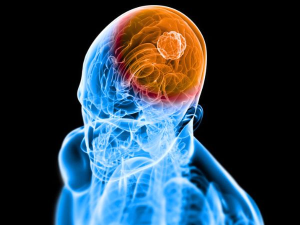
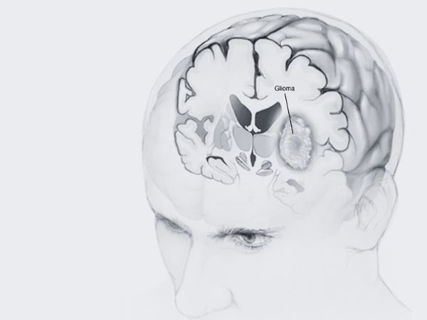
Glial cells in the brain or spinal cord are the source of gliomas, a form of tumor. The supporting cells of the nervous system, glial cells give neurones structure, defence, and nourishment. Roughly 80% of all malignant brain tumors and 30% of all brain tumors are gliomas.
The type of glial cell from which gliomas originate determines their classification:
- (from astrocytes) Astrocytomas
- (from oligodendrocytes) Oligodendrogliomas
- From ependymal cells, ependymomas
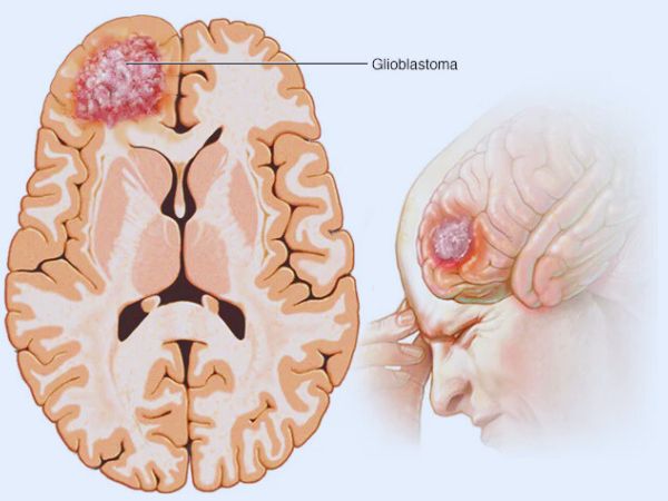
Furthermore, gliomas are categorised by the World Health Organisation (WHO) into four categories (I to IV) according to how aggressive they appear under a microscope. High-grade gliomas (Grades III and IV) grow more quickly and are more invasive than low-grade gliomas (Grades I and II), which grow more slowly. The most frequent and aggressive malignant primary brain tumor is glioblastoma multiforme (GBM), a Grade IV glioma.
Do gliomas come in different types?
Based on the kind of glial cell they originate in, gliomas can be divided into three primary categories. Multiple cell types can be found in certain gliomas. These are known as mixed gliomas by medical professionals. Based on their growth rate and other characteristics, they classify each form of glioma as low-, mid-, or high-grade.
Among the gliomas are:
- Glioblastomas and diffuse intrinsic pontine gliomas (DIPGs) are examples of astrocytes: Astrocytes are the cells that give rise to these tumors. Astrocytomas that grow quickly or are extremely aggressive are called glioblastomas. In adults, they are the most prevalent malignant brain tumor. Children frequently develop gliomas called astrocytes. DIPG is an uncommon but extremely aggressive kind of childhood brain cancer. It primarily affects children and develops in the brain stem.
- Ependymomas: These tumors originate in glial cells called ependymocytes. Ependymomas typically develop in the brain’s or spinal cord ventricles. They do not spread outside of the brain or spine, but they may do so through the cerebrospinal fluid, which envelops and shields the brain and spinal cord. About 2% of all brain tumors are ependymomas. Children are more likely than adults to have them.
- Oligodendrogliomas: These tumors begin as oligodendrocytes, which are glial cells. Although oligodendrogliomas typically grow more slowly, they have the potential to become more aggressive over time. They hardly ever spread outside of the brain or spine, just like ependymomas. Adults are more likely than children to have them. One to two percent of all brain tumors are oligodendrogliomas.
Glioma symptoms
The location, size, and growth rate of the tumor all affect the symptoms of gliomas. Typical signs and symptoms include:
- A headache
- Seizures
- Changes in personality or cognition
- Issues with memory
- Sensory abnormalities or motor weakness
- Visual disruptions
- Dizziness
- Weakness or numbness on one side of the body
- Difficulty in keeping balance and walking.
- Vomiting & Nausea
Who is susceptible to gliomas?
Gliomas can happen to anyone, but the following things could make you more susceptible:
- Age: Children under the age of twelve and older individuals over 65 are most likely to develop gliomas.
- Ethnicity: Compared to other races, white people may be more susceptible to gliomas.
- Family history: You may be more susceptible to gliomas if you have certain inherited genetic problems.
- Sex: Men are slightly more likely than women to get gliomas.
- Exposure to radiation or toxins: Prolonged or recurrent exposure to radiation or certain chemicals may raise your risk.
What is the frequency of gliomas?
In the United States, about 80,000 people receive a new primary brain tumor diagnosis every year. Gliomas make up about 25% of these.
Gliomas, especially those of the central nervous system (CNS), are seen in 5–10 out of every 100,000 people in India. About 2% of all malignant neoplasms in India are brain tumors, making up a sizable part of all brain tumors. Astrocytomas are a common kind of glioma, which can occur in both children and adults.
Why do gliomas occur or what is the cause?
According to research, gliomas and other brain and spinal cord tumors are caused by alterations in DNA. DNA is found in our genes. They guide cells on how to divide and multiply. Cells can proliferate uncontrollably if our genes’ DNA undergoes mutations.
Parents may pass on genetic mutations to you. They can also happen all at once while you’re alive.
What side effects might gliomas cause/complications?
The following are glioma complications that could be fatal:
- Bleeding in the brain, or brain haemorrhage.
- A brain herniation occurs when brain tissue shifts from its typical location within the skull.
- Hydrocephalus, or accumulation of fluid in the brain.
- Pressure within your head.
- Seizures.
How can gliomas be identified or diagnosed?
In addition to reviewing your medical history, your healthcare professional assesses your symptoms. A thorough neurological and physical examination will also be performed.
The most popular imaging tests for brain tumors are MRIs and CT scans. In addition to other tumors in your body, your doctor searches for brain tumors.
Doctor will perform a biopsy if your imaging scans reveal an abnormal lump. The process of analysing a tissue sample is called a biopsy. The biopsy will assist them in figuring out:
- If the growth is malignant.
- If an aberrant gene is the cause of the tumor.
- The tumor’s cell structure.
- The tumor’s grade, or aggressiveness.
Care and Therapy
How do you cure a glioma?
The following variables will affect your glioma treatment plan:
- If you have already had treatment for brain cancer.
- The tumor’s size, type, and location.
- Your age.
- Your well-being.
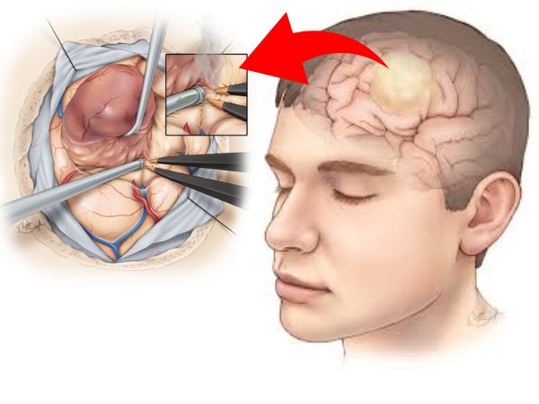
Surgery is typically the first course of treatment for gliomas. If the tumor is easily accessible, a surgeon might be able to remove it altogether. However, gliomas can be challenging to fully eradicate, particularly if they are in close proximity to sensitive brain regions or are difficult to reach.
Surgery should be followed by other therapies like radiation therapy and chemotherapy. These are adjuvant therapy, which means that following surgery, they eliminate any cancer cells or tumor components that may still be present. However, your doctor may employ radiation therapy or chemotherapy as your main therapies if a tumor cannot be removed.
Operation/Surgery
The most popular procedure for glioma removal is an open brain surgery called a craniotomy. You might be a candidate for laser ablation, depending on the tumor’s size and location. In this minimally invasive procedure, a brain tumor can be completely or partially destroyed using laser heat.
To guide the procedure, a surgeon may employ specialised methods like brain mapping or imaging. Brain mapping reveals the parts of your brain that regulate essential processes. By knowing this, your surgeon can prevent damaging or destroying vital brain tissue.
Radiation
Strong radiation doses are used in radiation therapy to eradicate tumors. For gliomas, your doctor might suggest radiation treatment. By precisely targeting the tumor’s form, radiation therapy reduces the possibility of harming nearby tissues.
Brachytherapy is another type of radiation treatment that you could get. To treat a tumor, a medical professional utilises radiation sources near the tumor. Radiation is released from the sources without damaging surrounding tissues.
Chemotherapy
Chemotherapy is the process of killing cancer cells with chemicals. It cures a variety of cancers. This treatment can be administered directly or orally.
One frequent chemotherapeutic medication used to increase the effectiveness of radiation therapy is temozolomide.
Avoidance/Prevention
How can gliomas be avoided?
The majority of glioma risk factors, including age and race, are uncontrollable. However, the evolution of low-grade gliomas into high-grade gliomas may be slowed or stopped by early detection and treatment. You might wish to think about genetic testing if brain tumors run in your family. Discuss the advantages and disadvantages of genetic testing with a genetic counsellor or your healthcare provider.
Additionally, it is wise to:
- Don’t expose your head to too much radiation.
- Keeping up a healthy way of living.
Prognosis and Outlook
What is the prognosis for glioma patients?
Glioma survival rates differ depending on the kind, grade, and age of the tumor. The prognosis may also be impacted by specific mutations. The prognosis is poorer for those who are diagnosed and treated later in life. Adults and children with low-grade ependymomas, oligodendrogliomas, and astrocytomas have the highest five-year survival rates. For glioblastomas, it is the lowest (between 6% and 20%).
Glioma Gamma Knife Radiosurgery
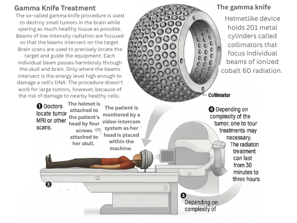
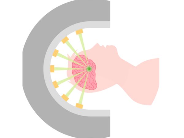
A type of stereotactic radiosurgery (SRS) called “gamma knife” uses intensely concentrated radiation beams to target the tumor while exposing the least amount of healthy brain tissue possible. It is especially helpful for gliomas that are in sensitive brain regions or are difficult to physically approach.
Radiosurgery with Gamma Knife for Low-Grade Gliomas
Gamma Knife radiosurgery provides a less invasive treatment option for low-grade gliomas (WHO Grade I and II) that can:
- After partial resection, control the growth of any remaining tumor.
- Treat well-defined, tiny lesions without requiring open surgery.
- Preserve neurocognitive function by delaying or minimising the need for whole-brain radiation.
Research has demonstrated that Gamma Knife can successfully treat low-grade gliomas with few adverse effects, providing long-lasting local control and enhanced quality of life. Younger patients or those with tumors in functionally sensitive areas benefit most from it.
Radiosurgery with Gamma Knife for High-Grade Gliomas
Treatment for high-grade gliomas, such as glioblastoma multiforme (GBM), is difficult and aggressive. Uses for Gamma Knife radiosurgery include:
- Recurrent High-Grade Gliomas: Used as a salvage treatment when tumors return following initial chemotherapy, radiation, and surgery.
- Targeting isolated or satellite lesions that are not responsive to additional surgical procedures is the goal of small residual or metastatic lesions.
In certain situations, Gamma Knife can effectively control tumors, extending survival and improving quality of life, however it should not be used in place of conventional treatment methods. To maximise results, multidisciplinary planning and careful patient selection are essential.
Gamma Knife Radiosurgery Benefits
- Precision: Accurately targets tumors within millimetres.
- Safety: Reduces the amount of radiation that reaches healthy brain tissue.
- Convenience: A short recovery period following an outpatient treatment.
- Repeatability: For recurrent tumors, it is possible to repeat if required.
In conclusion
Glioma treatment necessitates a customised, interdisciplinary strategy that strikes a balance between tumor control and neurological function preservation. A potent weapon in this arsenal is Gamma Knife radiosurgery, which gives patients with both low-grade and high-grade gliomas hope and better results.
For the best outcome, it is essential to speak with a skilled neurosurgeon who can explain the various treatment choices, including cutting-edge methods like Gamma Knife radiosurgery, if you or a loved one has been diagnosed with a glioma.
Sources:
- https://pmc.ncbi.nlm.nih.gov/articles/PMC4991137/
- https://www.ncbi.nlm.nih.gov/books/NBK441874/
- https://www.ncbi.nlm.nih.gov/books/NBK441874/#_article-18547_s13_
- https://my.clevelandclinic.org/health/diseases/21969-glioma
- https://www.mayoclinic.org/diseases-conditions/glioma/symptoms-causes/syc-20350251
- https://en.wikipedia.org/wiki/Glioma
- https://www.cancerresearchuk.org/about-cancer/brain-tumors/types/glioma-adults
- https://www.hopkinsmedicine.org/health/conditions-and-diseases/gliomas
- https://www.cancer.gov/publications/dictionaries/cancer-terms/def/glioma
- https://braintumorresearch.org/pages/types-of-brain-tumors-glioma?srsltid=AfmBOoqH0Fueb1M3QbNjKjiS2AYdxR-3CORqtBkctpVf9LrxEQ9I_Hzy