A SYSTEM AND METHOD FOR AUTOMATED MIDLINE SHIFT QUANTIFICATION IN BRAIN CT IMAGES USING DEEP LEARNING
Overview
The brain may occasionally shift from its typical central location inside the skull as a result of a brain injury. We refer to this process as midline shift (MLS). Midline shift may seem like a straightforward measurement, but it can be a crucial sign of brain swelling, haemorrhage, mass effect, and rising intracranial pressure.
Doctors frequently assess a CT scan in patients with traumatic brain injury (TBI) to ascertain whether midline displacement is present and, if so, how serious it is. Traditionally, a radiologist or neurosurgeon may need to manually measure and carefully evaluate the results.
Automatically detecting and measuring midline displacement from a brain CT scan is becoming possible because to developments in artificial intelligence (AI), deep learning, and medical image analysis.
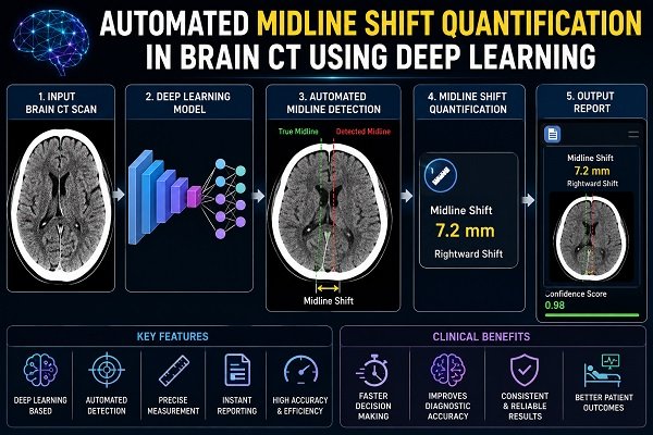
A deep learning-based automated midline shift quantification system and method may be able to analyse CT scans, identify key anatomical features, detect midline displacement in the brain, and produce a quantitative estimate to help physicians.
The potential of deep learning-based methods for automated midline shift identification in traumatic brain injury has been investigated by research led by Dr. Deepak Agrawal and colleagues. The Indian Journal of Neurotrauma published a study in 2024 that looked at 15 studies that used AI-based methods to identify and measure midline displacement. The review emphasised the potential of these technologies as well as their present drawbacks.
Midline Shift: What Is It?
Normally, the brain is located roughly symmetrically inside the skull. The midline of the brain can be described using an imaginary middle line.
When anything takes up extra space inside the skull, like:
- A traumatic haemorrhage in the brain
- Haematoma subdural
- Haematoma in the epidural space
- Swelling of the brain
- Tumour in the brain
- Big stroke
- Abscess
- Other lesions that take space
It is possible to push the surrounding brain tissue out of its natural place.
Midline shift is the term for this displacement.
Why is midline shift important?
The structure of the skull is inflexible. Its ability to handle more volume is restricted. Brain tissue may be crushed and displaced when bleeding or edema raises intracranial pressure.
Therefore, a large midline shift may be a symptom of a severe mass impact and may be linked to a major decline in neurological function.
Crucially, midline shift should not be perceived in a vacuum. In addition to the patient’s age, injury mechanism, neurological examination, CT results, and other clinical criteria, doctors take this into account.
How Is Midline Shift Measured Conventionally?
The radiologist or neurosurgeon determines the proper anatomical landmarks on a CT scan and calculates the extent to which the brain’s structures have deviated from their anticipated central location.
The procedure could entail:
- Examining several CT slices.
- Determining which anatomical structures are pertinent.
- Figuring out the usual or expected midline.
- Determining which brain structures are shifted.
- Calculating the separation between the displaced structures and the reference midline.
- Interpreting the measurement in conjunction with further CT results.
Expertise and careful image interpretation are needed for this.
By enabling a computer algorithm to methodically examine CT data, automated image analysis seeks to lessen some of this manual labour.
Automated Midline Shift Quantification: What Is It?
The use of computer methods to determine whether midline displacement is present and calculate its amount from brain CT scans is known as automated midline shift quantification.
A deep-learning system can be trained to identify patterns in CT scans linked to normal anatomy and anatomical displacement rather than depending solely on manual measurement.
Replacing radiologists and neurosurgeons is not the ultimate objective. Instead, aim is to offer a screening and measurement tool with AI assistance that can facilitate quick and impartial evaluation.
How Is Deep Learning Beneficial?
Neural networks are used in deep learning, a subfield of artificial intelligence, to extract patterns from massive volumes of data.
These networks can learn to identify intricate visual features in medical imaging that would be challenging to characterise using straightforward mathematical principles.
A deep learning model for brain CT analysis might pick up characteristics related to:
- Anatomy of the brain
- Ventricular anatomy
- Boundaries of the skull and the brain
- Anatomical landmarks
- Asymmetry of the brain
- Mass effect and bleeding
- Midline structure displacement
The system can analyse a fresh CT scan and produce an automatic evaluation when it has been properly trained and certified.
How Would an Automated Midline Shift System Operate?
There are multiple levels to understanding a reduced workflow.
Step 1: Acquisition of CT Scans
A typical non-contrast head CT scan, which is frequently utilised in the initial assessment of traumatic brain damage, is performed on the patient.
Step 2: Processing Images
The AI system can analyse the CT pictures after they have been processed.
When necessary, the program may normalise the images and locate pertinent anatomical regions.
Step 3: Identification of Anatomical Features
The deep learning model finds significant anatomical landmarks or features that aid in determining the predicted midline of the brain.
Step 4: Identification of the Midline
Based on data from the CT scans, the system calculates the patient’s anatomical midline.
Because severe brain injuries can affect normal anatomy, this is especially crucial.
Step 5: Displacement Detection
Whether brain structures have shifted from the estimated midline is assessed by the algorithm.
Step 6: Measurement
The system can calculate the amount of displacement if a shift is observed, possibly representing it in millimetres.
Clinical Visualisation in Step Seven
The outcome might be shown on the CT scan as a measurement, annotation, or visual overlay by an AI-assisted system.
Step 8: Clinician Evaluation
The qualified healthcare professional has the final say in the interpretation, taking into account both the automated outcome and the full clinical and radiological picture.
What Makes 3D Deep Learning Crucial?
While individual CT slices are two-dimensional, the anatomy of the brain is three-dimensional.
This poses a problem since a structure that looks displaced on one slice could look different on another.
By analysing data from several CT slices, a 3D convolutional neural network (3D CNN) may be able to identify spatial correlations throughout the whole volume of the brain.
A 3D CNN-based automated screening method was created in a study by Dr Agrawal and colleagues utilising 176 head CT scans from traumatic brain injury patients. Twenty scans were used for testing and 156 scans were used for training. With stated sensitivity of 40%, specificity of 70%, and overall accuracy of 55%, it accurately detected 7 out of 10 midline-shift instances and 4 out of 10 non-midline-shift cases in the test set. The authors stressed the need for more improvement and validation while concluding that automated screening demonstrated viability.
These findings are significant because they show the potential of AI-based midline shift screening as well as its present limitations.
Why Is Automated Midline Shift Detection Challenging?
Measuring midline displacement may appear simple at first. In fact, after a serious injury, the brain can become severely damaged.
Automated measuring may face a number of difficulties.
- The intricate structure of the brain: It is challenging for an algorithm to determine a universal midline since various patients have different anatomical variances.
- Serious anatomical abnormality: Normal structures can be severely distorted by large haematomas and oedema.
- Various CT scanners: Depending on the brand of the scanner, the acquisition method, and the image quality, CT images can differ.
- Minor adjustments: High measurement precision is necessary to detect a very little displacement.
- Big datasets are necessary: For robust performance, deep learning models often need diverse and adequately structured data sets.
- Various clinical manifestations: It’s possible that a model that was mainly trained on one kind of TBI won’t function as well in other populations.
- The requirement for outside verification: When evaluated at a different hospital or with different CT methods, a model may perform differently even though it performed well on its development dataset.
Agrawal, Joshi, and Poonamallee’s 2024 assessment found significant differences in published AI approaches, datasets, and evaluation techniques. The stated sensitivity and specificity ranged from 70% to 100% and 73% to 97.4%, respectively, among the analysed research, demonstrating why findings from various algorithms cannot be simply compared without taking into account their techniques and datasets.
Why Could AI-Assisted Midline Shift Detection Be Beneficial?
- Quicker evaluation: Time is of the essence in neurosurgical emergencies. Automated analysis could quickly identify facts that could be important.
- Measuring objectively: An automated numerical measurement could lessen measurement variability and supplement visual interpretation.
- Assistance to emergency teams: In high-volume emergency settings, AI-based screening may be able to help prioritise CT scans that need immediate evaluation.
- Quantitative data: Rather of merely classifying a change as “present” or “absent,” automated methods may be able to offer quantifiable data.
- Tracking modifications: Clinicians may someday be able to compare imaging tests over time with the aid of quantitative data.
- Potential for decision-support: Clinical decision-support systems may benefit from automated assessments when paired with additional CT and clinical data.
But rather than being an autonomous decision-maker, AI should be seen as an auxiliary technology.
Uses Other Than Traumatic Brain Injury
Midline shift can happen in a number of neurological diseases, even though traumatic brain injury is a significant application.
The following are some possible uses for automated midline shift analysis:
- Brain damage caused by trauma
- Haemorrhage within the brain
- Haematoma subdural
- Haematoma in the epidural space
- Cerebral oedema following a large ischaemic stroke
- Brain tumours
- An abscess in the brain
- Other circumstances that have a large mass effect
For a number of TBI-related anomalies, such as haemorrhage, haematoma volume, and midline displacement, automated CT analysis is being investigated more and more.
From Identification to Quantification: The Significance of Measurement
The contrast between identifying and quantifying midline displacement is crucial.
Identification
The system responds:
“Is there evidence of midline shift?”
Quantification
The system tries to respond to:
“If there is a shift, approximately how large is it?”
Compared to a straightforward yes/no classification, quantification may provide additional information.
An automated system might, for instance, report:
Yes, there was a midline shift.
X mm is the estimated displacement.
A skilled doctor must next assess the measurement’s true clinical relevance in light of the patient’s overall health.
Approaches Based on Symmetry vs. Landmarks
Numerous methods have been employed in the study of automated midline shift detection.
Methods Based on Symmetry
By examining the symmetry of the brain, these techniques strive to determine the natural midline.
The underlying idea is very obvious: the brain’s usual symmetry may be disrupted if one side of the brain is displaced.
Methods Based on Landmarks
These methods pinpoint particular anatomical features that can be used as benchmarks.
These structures’ expected locations can be estimated by the algorithm and compared to their actual locations.
Both symmetry-based and landmark-based methods are included in the literature examined by Agrawal and colleagues, with deeper neural networks being used in subsequent systems.
AI’s Role in Emergency Neurosurgery
Making quick decisions is often necessary in emergency neurosurgery.
A patient could show up with:
- Diminished awareness
- Vomiting and a severe headache
- Weakness
- Poor pupils
- Seizures
- Confusion
- A history of severe head injuries
Haemorrhage, oedema, and mass effect can all be seen on a CT scan.
An additional layer of support for physicians could be provided by an automated AI system that promptly identifies probable midline displacement.
Automation is not the only goal. Enhancing the speed, consistency, and quantitative interpretation of clinically significant CT results is the goal.
What Are Automated Midline Shift Quantification’s Drawbacks?
Before being used in routine clinical decision-making, AI in healthcare needs to be thoroughly validated.
Insufficient training data
The entire range of patients and imaging situations may not be fully represented by a model that was developed on a very limited dataset.
The ability to generalise
When the model is applied in a different hospital, nation, patient population, or CT scanner, performance may vary.
False negatives and false positives
Sometimes an AI system fails to detect an actual anomaly or detects a shift that is not clinically significant.
Anatomical intricacy
The brain can be severely distorted by severe trauma, which makes automated midline assessment difficult.
Absence of a uniform assessment
The datasets, definitions, algorithms, and performance metrics used in various studies vary. Standardisation and additional validation were especially noted in the 2024 review as crucial areas for further study.
Clinical judgement cannot be replaced by AI.
One part of the patient evaluation process is the CT scan. Clinical history, neurological examination, and other studies are still crucial.
Automated Brain CT Analysis’s Future
AI-assisted neuroimaging is probably going to go beyond identifying one anomaly at a time in the future.
A thorough AI system would be able to examine a head CT scan for several results at once, including:
- Haemorrhage within the brain
- Type of haematoma
- Volume of haematoma
- The midline shift
- Swelling in the brain
- Compression of the ventricles
- Changes in the basal cistern
- Fractures of the skull
These tools might help radiologists and neurosurgeons by offering a structured quantitative summary.
Research has previously shown that deep learning may be used to automatically quantify a number of CT characteristics associated with acute traumatic brain damage.
Larger datasets, multi-center validation, standardised performance evaluation, and cautious incorporation into actual clinical workflows are all necessary for the next phase.
How Will Patients Be Affected by This Innovation?
The technique is easy for a patient or family member to understand:
The images are obtained from a CT scan. AI aids in the analysis of those pictures. The clinical choice is made by the physician.
One possible advantage is that significant anomalies like midline displacement could be found and recorded more rapidly and reliably.
This could be especially helpful in emergency situations where medical professionals have to quickly assess the extent of a brain lesion and determine whether immediate action is required.
However, healthcare experts with the necessary training should always interpret measurements produced by AI.
AI-Powered Neurosurgical Innovation and Dr. Deepak Agrawal
Research on the nexus of neurosurgery, artificial intelligence, and medical imaging has been conducted by Dr. Deepak Agrawal of the Department of Neurosurgery at the All India Institute of Medical Sciences (AIIMS), New Delhi.
His research on 3D convolutional neural networks for the analysis of head CT scans in traumatic brain injury is part of his work on automated midline shift identification.
Automated Midline Shift Detection and Quantification in Traumatic Brain Injury: A Comprehensive Review, which he co-authored in 2024, looked at the development of AI-based techniques and highlighted the potential of deep learning while acknowledging the need for more validation and standardisation.
In conclusion
Deep learning-based automated midline shift measurement is a significant nexus of medical imaging, artificial intelligence, and neurosurgery.
When it comes to the severity and mass effect of brain lesions, midline shift can offer important information. It may be possible to make CT interpretation quicker, more repeatable, and more quantitative by automating its detection and measurement, especially in emergency situations.
Automated midline shift screening is technically possible, according to research using deep learning and 3D CNN techniques. However, current research indicates that before such systems can be regularly relied upon, larger datasets, multi-center validation, standardised evaluation techniques, and robust clinical testing are necessary.
Clinical knowledge and intelligent technology will work together more and more in neurosurgery in the future. AI is unlikely to take the role of neurosurgeons; rather, it might be most useful in giving medical professionals quicker, more objective, and useful information when every second counts.
FAQ’s
1. What is the brain’s midline shift?
The movement of the brain’s core structures from their typical location is known as a midline shift. It can happen when pressure is created inside the skull by bleeding, swelling, a tumour, or another space-occupying lesion.
2. Is the midline shift risky?
A large midline shift may be linked to severe neurological damage and may be an indication of major pressure or mass influence on the brain. The degree of change and the patient’s general clinical status determine its relevance.
3. How is midline shift identified?
In acute neurological emergencies, it is typically detected on brain imaging, especially CT scans. After analysing the pictures, a radiologist or neurosurgeon may calculate the displacement.
4. What does automatic midline shift detection entail?
This AI-based method determines whether midline displacement is present by analysing brain CT scans.
5. What is quantification of automated midline shifts?
By calculating the amount of displacement—typically expressed as a distance in millimeters—quantification goes one step farther.
6. How is midline shift measured using deep learning?
Labelled CT scans can be used to train deep learning algorithms to identify anatomical patterns and structures related to midline displacement. Information from several CT slices can be analysed by sophisticated models, such as 3D convolutional neural networks.
7. Can AI take the role of a neurosurgeon or radiologist?
No, AI ought to be regarded as a screening or clinical decision-support technology at this time. Qualified healthcare professionals should make the final judgements regarding interpretation and treatment.
8. Is it possible to employ automated midline shift detection in cases of traumatic brain injury?
It has a lot of potential for TBI research. Research has shown that deep learning can be used to screen for midline shift, but before any particular system can be deemed generally reliable for clinical usage, more validation is needed.
9. Does a midline shift always necessitate surgery?
No, surgery is not always required just because there is a midline shift. The underlying reason, degree of mass effect, neurological condition, imaging results, and other clinical considerations all influence treatment.
10. For acute brain injury, which CT scan is most frequently used?
When evaluating acute traumatic brain damage, a non-contrast head CT scan is frequently utilised.
11. Can AI identify further brain CT abnormalities?
Indeed. Research has studied AI for identifying and measuring intracranial hemorrhage, hematoma volume and other CT features associated with traumatic brain injury.
12. Describe a 3D CNN.
One kind of deep learning model that can analyse three-dimensional picture data is a 3D convolutional neural network. Instead of considering each image entirely separately, this can enable the algorithm in brain CT to take into account relationships between several CT slices.
13. How precise is automated midline shift measurement?
There are significant differences in accuracy between datasets and algorithms. Although published research shows encouraging results, direct comparisons are challenging due to methodological discrepancies. Additional multi-center, large-scale validation is required.
14. How will AI-based brain CT analysis develop in the future?
The long-term objective is to create dependable systems that can quickly identify and measure a variety of clinically significant anomalies and provide this data to physicians as decision-support.
Source: https://www.thieme-connect.com/products/ejournals/pdf/10.1055/s-0043-1777676.pdf