We are proud to share a major milestone in medical innovation and research! The Patent Office, Government of India, has officially granted a Certificate of Registration of Design (Design No: 517979-001, Class 24-01) for the 3D-PRINTED MICROFLUIDIC LIVE-CELL IMAGING DEVICE, patented in the name of Dr. Deepak Agrawal and his research team.
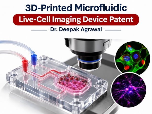
Official Patent & Design Registration Details
- Title of Design / Innovation: 3D-PRINTED MICROFLUIDIC LIVE-CELL IMAGING DEVICE
- Design No.: 517979-001
- Class: 24-01
- Serial No.: 259373
- Date of Registration: 16/09/2026
- Date of Issue: 28/10/2026
- Inventors / Patentees: Dr. Deepak Agrawal, Aparna Sharma, Dr. Prabhakar Singh, Nilesh Srivastava
- Issuing Authority: The Patent Office, Government of India
About the Innovation: 3D-Printed Microfluidic Live-Cell Imaging Device
Live-cell imaging plays a pivotal role in understanding cellular behavior, real-time drug response, and disease progression. This breakthrough aligns closely with modern advancements in study the brain before surgery and cutting-edge laboratory research methodologies like brain organoids and auspatial radiation mapping culture dish.
Traditional microfluidic platforms often face severe limitations regarding high manufacturing costs, lack of customizability, and complex fabrication workflows. This newly patented 3D-printed device overcomes these hurdles by integrating additive manufacturing with microfluidic precision, allowing researchers to observe dynamic cellular changes with high-resolution clarity under tightly controlled microenvironments. Furthermore, understanding complex cellular environments helps medical professionals better evaluate conditions related to neurodegenerative diseases and complex brain cancer types, symptoms, and causes.
Key Features and Technical Advantages
- High Precision Real-Cell Tracking: Enables detailed, continuous visualization of cellular interactions and morphological changes.
- Cost-Effective Prototyping: Utilizing 3D-printing technology drastically reduces production costs and lead times for modern laboratory setups.
- Versatile Applications: Highly beneficial for neurology, oncology, high-throughput drug screening, and fundamental tissue engineering.
Impact on Medical Science and Neurosurgical Research
By leveraging advanced 3D-printing techniques, this design allows rapid prototyping and low-cost manufacturing, making advanced diagnostic tools more accessible for research across various disciplines. Bridging the gap between engineering and clinical translation, this indigenous innovation supports the vision of advanced healthcare technology development in India.
The successful registration of this design marks a proud moment for indigenous research at AIIMS and sets a strong precedent for future medical device development. Staying at the forefront of medical technology also ties into advanced clinical treatments such as gamma knife radiosurgery and precision brain tumor removal surgery.
Future Directions and Clinical Translation
As we move forward, the integration of 3D-printed microfluidics into everyday clinical and laboratory workflows will open up new horizons in personalized medicine. Researchers will be able to test targeted therapies on live tissue models efficiently before taking surgical or therapeutic decisions.
Stay tuned to Dr Deepak AIIMS for more updates on medical patents, technological breakthroughs, and advanced neurosurgical innovations.