AuSpatial Radiation Mapping Culture Dish: A New Approach to Spatially Mapping Gamma Knife Radiation Effects
Knowing how to precisely identify, monitor, and investigate radiation-induced alterations in cells, spheroids, and organoids using a 3D-printed grid-based culture plate
Medical & Research Disclaimer: This article reviews a research methodology (AuSpatial Radiation Mapping Culture Dish) developed for laboratory research in radiobiology and neuro-oncology. It is not a clinical treatment device or a substitute for professional medical care.
Highly concentrated radiation can be delivered to precisely defined targets using contemporary radiation technologies like Gamma Knife radiosurgery. But examining the biological impacts of this highly localised radiation in a lab setting poses a distinct problem: how can scientists pinpoint the precise area that was exposed to radiation and then locate that same area under a microscope?
A customised 3D-printed, grid-based confocal culture dish is used in a research methodology created by Dr. Deepak Agrawal and associates at AIIMS, New Delhi, to address this problem. The method makes it possible to correlate radiation-targeted areas with later microscopic measurements by introducing a fixed spatial coordinate system into the culture environment.
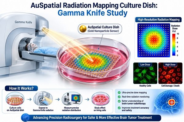
This unique idea, referred to here as an AuSpatial Radiation Mapping Culture Dish, may be especially helpful for translational research, radiation biology, organoid research, and neuro-oncology.
An AuSpatial Radiation Mapping Culture Dish: What Is It?
A specialised culture dish with an exact spatial grid is called an AuSpatial Radiation Mapping Culture Dish.
This grid separates the culture region into distinguishable pieces, in contrast to a traditional cell-culture dish where researchers do not have a stable coordinate reference. Thus, a cell cluster, spheroid, or organoid’s location can be noted by researchers prior to radiation and then revisited during subsequent microscopic analyses.
The underlying study created a 5 × 5 grid with indexed square cells that were roughly 2 mm × 2 mm in size. The grid’s overall dimensions were 20 mm by 20 mm, and its ultrathin thickness was about 0.2 mm.
To put it simply, it functions similarly to covering a biological sample with a map grid.
Rather than stating: “The radiation affected this general area.”
Researchers can try to determine: “The radiation affected this specific indexed region, and this is the same region we are examining under the microscope.”
Why Does Radiation Research Need Spatial Mapping?
The purpose of gamma knife technology is to target a specific area with concentrated radiation. The problem occurs when scientists wish to investigate the effects of exposure on individual cells or three-dimensional biological models.
Dishes from traditional cultures typically don’t offer a fixed coordinate system. Moving the precise area that was checked before to radiation can therefore be challenging.
When studying, this becomes very crucial:
- Organoids of the brain
- Spheroids of tumours
- Cell groups
- Tissue models in three dimensions
- Cellular damage caused by radiation
- Sensitivity to radiation
- Resistance to radiation
- Neurotoxicity
The lack of spatial reference markers, which makes it challenging to properly correlate radiation delivery sites with subsequent confocal microscopy data, is identified in the research paper as a significant disadvantage.
How Does the Grid-Based Culture Dish Operate?
Three significant technologies are combined in this concept:
1. Spatial Grid Printed in 3D
Fused deposition modelling (FDM) 3D printing is used to create the grid once it has been developed using computer-aided design (CAD) software.
Two materials were assessed in the study: PLA (polylactic acid) and PETG (polyethylene terephthalate glycol).
The grid can be tailored to the requirements of a specific experiment thanks to the usage of 3D printing.
2. Radiation from Gamma Knives
A certain grid region can be chosen as the intended radiation target, and cells or organoids are placed inside the indexed compartments.
The researchers can record the targeted site and re-identify it after irradiation since the grid offers a fixed coordinate system. Advances in treatment protocols are frequently benchmarked against techniques like Gamma Knife, Proton Beam, or CyberKnife.
3. Confocal Microscopy
Confocal microscopy is used by researchers to study biological alterations following irradiation.
During follow-up imaging, it is feasible to return to the same area repeatedly thanks to the indexed compartments.
Researchers can use this to examine changes like:
- Morphology of cells
- Apoptosis
- Nuclear disintegration
- Disruption of the structure
- Migration of cells
- Increase of reactive oxygen species
- Organoid structural alterations
What Is Unique About the Grid?
The grid was intended to be extremely thin, perhaps 0.2 mm.
This is significant because materials that are positioned between biological material and a radiation source have the ability to affect radiation by absorption, scattering, or attenuation.
As a result, the platform was created by the researchers to balance two requirements: low interference with radiation and imaging combined with mechanical stability. Preventing adverse radiation effects is critical when engineering tissue-dish platforms.
According to the study, the manufactured grids remained compatible with confocal microscopy and retained their structure throughout handling, sterilisation, irradiation, and imaging.
How Do You Determine the Grid Coordinates?
“X,” “Y,” and “S” markings, as well as an indexing hole, are among the orientation and indexing elements included on the platform.
During radiation therapy and microscopy, these markers assist researchers in maintaining a constant orientation.
This seemingly straightforward function is crucial since it can be challenging to compare images captured at different times due to even little positioning variations.
Is It Possible to See the Grid While Imaging?
Indeed.
The study’s key discovery was that confocal microscopy and cone-beam computed tomography (CBCT) could both be used to visualise the grid.
While confocal microscopy enabled researchers to recognise the same indexed compartments during biological investigation, CBCT visualisation enabled the grid to function as a spatial reference during the irradiation/localization operation.
This establishes a link between:
Radiation location → Grid coordinate → Microscopic field → Biological response
The system’s core value is that correlation.
What Can This Technology Be Used for Research?
1. Cellular Damage Caused by Radiation
Numerous cellular effects can be caused by radiation. Researchers can look at changes in specific areas following radiation exposure using spatially indexed imaging.
2. Organoids of the Brain
Aspects of the structure of the human brain can be replicated in three-dimensional laboratory models called brain organoids, similar to how mini-brains grow from fetal brain tissue.
According to the research, the platform may be helpful in examining the effects of localised radiation on brain organoids, including modifications pertaining to inflammation, apoptosis, DNA damage, and other biological reactions. Understanding these effects also provides insight into brain organoids in traumatic brain injury research.
3. Spheroids of Tumours
Tumour biology and treatment response can be investigated using tumour spheroids.
Tumours may respond differently to radiation in different areas because to the presence of regions with varying oxygen levels, metabolic states, and proliferative activity. Therefore, spatial mapping could aid researchers in examining this heterogeneity across various types of cancer.
4. Radiation Resistance and Sensitivity
Radiation can affect tumour locations and different tissues in different ways.
Researchers may be able to better understand why some areas are more radiation-sensitive or resistant by using a spatially indexed culture platform.
5. Longitudinal Research
Repeated imaging over time may be one of the most practical uses.
Researchers can compare changes across several time periods by going back to the same indexed compartment before and after radiation, enabling teams to effectively study the brain before surgery or interventions.
What Function Does 3D Printing Serve?
Researchers can make lab equipment that is precisely tailored to their experimental needs thanks to 3D printing.
Rather than depending solely on products that are sold commercially, researchers can alter:
- Grid dimensions
- Dimensions of the compartment
- Thickness of the grid
- The quantity of compartments
- Markers of orientation
- The overall geometry
The study identifies cost-effectiveness, quick prototyping, and customisation as key benefits of FDM-based manufacturing.
This adaptability may be especially helpful when various biological models call for various spatial arrangements.
Possible Function in Neuro-Oncology
Translational neuro-oncology research may benefit from this idea.
Clinically, Gamma Knife is used to treat certain neurological diseases using highly targeted radiation, such as evaluating the role of Gamma Knife in glioblastoma, managing Gamma Knife in cavernous hemangioma, or administering Gamma Knife for giant brain tumors and Gamma Knife for uveal melanoma. However, suitable experimental models are needed to comprehend the biological reaction to localised radiation.
Researchers may link biological alterations seen under high-resolution microscopy to the actual region of radiation exposure with the aid of a grid-based culture platform.
This could aid in the investigation of:
- Radiation response to brain tumours such as gliomas, atypical meningioma, or a threatening brain tumor
- Neurotoxicity caused by radiation
- Heterogeneity of tumours
- Sensitivity to radiation
- Resistance to radiation
- Research on organoid-based therapies
- Research on precision radiotherapy
The platform may be helpful for translational neuro-oncology research and organoid-based radiobiological studies, according to the researchers.
Could Personalised Medicine Benefit from This?
Maybe in the future.
More research is being done on patient-derived organoids as models to forecast how a patient’s tumour would react to therapy.
According to the study, researchers may someday be able to better understand individualised radiosensitivity and treatment responses by merging patient-derived organoids with geographical radiation mapping. But rather than being a proven therapeutic use, this is still a direction for future research.
It is crucial to stress that the AuSpatial radiation mapping culture dish is not a clinically proven therapy tool, but rather a research methodology.
What Benefits Are There?
Several possible benefits of the grid-based platform are identified by the research:
Accurate spatial localisation
- Researchers are able to pinpoint the radiation-exposed area.
Enhanced repeatability
- In further imaging sessions, the same coordinate may be revisited.
Radiation and microscopy are correlated
- Confocal microscopy fields and radiation coordinates can be directly compared.
Suitability for 3D biological models
- Organoids and other three-dimensional models can be utilised with the platform.
Personalisation
- The design may be altered for various experiments thanks to 3D printing.
Cost-effectiveness
- For laboratory prototype, FDM-based fabrication offers a practical method.
Monitoring over time
- It is possible to investigate the same area more than once over time.
What Are the Restrictions?
It’s crucial to avoid exaggerating the capabilities of this technology.
The study’s main objectives were structural validation and proof-of-concept. There was no quantitative dosimetric analysis done.
The authors clearly point out that more research is required to determine whether the grid material causes minute changes in the distribution of radiation exposure. Film dosimetry and Monte Carlo simulations were proposed as potential methods.
Other areas that need more research are as follows:
- Biological interactions over time with printed materials
- Material optimisation for extended live-cell research
- Automated registration of images
- Coordinate tracking that is automatic
- Combining AI-assisted spatial analysis with radiation dosimetry
Future enhancements like translucent grids, fluorescent fiducial markers, integrated dosimetry sensors, microfluidic integration, and AI-assisted spatial analysis are suggested by the study.
How Will the Future Turn Out?
The following could be combined in the future generation of spatial radiation mapping systems:
3D printing + organoids + radiation dosimetry + confocal microscopy + artificial intelligence
Future technologies might, for instance, automatically detect a radiation-exposed area, register it with microscopic images, and monitor cellular changes over time. Modern clinical developments are already witnessing artificial intelligence in Gamma Knife treatment workflows.
AI-assisted image analysis may be able to quantify:
- Modifications to the shape of cells
- Death of cells
- Damage to structures
- Reaction to radiation
- Variations in treatment sensitivity by region
These advances may improve the quantitative and repeatability of spatially resolved radiation biology.
What Makes This Innovation Significant?
The usage of a 3D-printed grid is only one aspect of this technology’s true significance.
It is the development of a shared spatial language between biological imaging and radiation delivery.
Researchers can determine the location of radiation delivery by using radiation treatment.
Researchers can learn about biological events through microscopy.
These two bits of information are connected by the grid.
This can make the question challenging: “Where did the radiation-induced alteration take place?”
Into a research question that is far easier to handle: “What biological alterations took place in this particular, predetermined area exposed to radiation?”
In intricate three-dimensional models like organoids, where biological reactions may differ from one area to another, this distinction can be especially helpful.
In Conclusion
The AuSpatial Radiation Mapping Culture Dish is a novel solution to one of the real-world problems in radiation biology: precisely linking the location of radiation delivery to the biological model’s subsequent outcomes.
The AIIMS research team’s 3D-printed grid-based confocal dish offers a fixed spatial reference that can be applied to confocal microscopy, CBCT localisation, and Gamma Knife irradiation. In cell and organoid research, the proof-of-concept study showed better spatial localisation, imaging compatibility, and structural stability. In broader experimental frameworks, research often utilizes biomarkers such as analyzing blood tests to predict brain changes after Gamma Knife exposure.
This approach shows how 3D printing, precision radiation technology, improved microscopy, and organoid models can be used to provide more repeatable and spatially precise radiation research, even though more dosimetric, biological, and clinical research is required.
Such technologies may eventually aid researchers in better understanding radiation response, tumour heterogeneity, and treatment resistance for the larger fields of neuro-oncology and translational neuroscience. They may also help develop precision radiotherapy in the future.
This study, which emphasises the possibility of integrating neurosurgery, biomedical engineering, imaging, and translational research to address challenging issues in contemporary neuroscience, was conceived and overseen by Dr. Deepak Agrawal, neurosurgeon at AIIMS Delhi.
Frequently Asked Questions (FAQs)
- What is a Culture Dish for AuSpatial Radiation Mapping?
Researchers may identify and consistently pinpoint precise locations exposed to localised radiation with the use of this specialised culture-dish platform that incorporates a 3D-printed indexed grid. - Is this a brand-new Gamma Knife procedure?
No. The technology’s main purpose is to serve as a platform for research on the effects of radiation on cells, spheroids, and organoids. Standard clinical Gamma Knife treatment is not altered or replaced by it. - What is a Gamma Knife?
Gamma Knife is a type of stereotactic radiosurgery that targets specific locations with high spatial precision by using highly focused radiation. The platform was created to aid in the investigation of localised radiation effects in lab models. - What makes a grid necessary?
Fixed spatial coordinates are typically not provided by a traditional culture dish. Researchers can pinpoint the same location both before and after irradiation because to the grid’s recognisable sections. - How does the grid appear?
A 5 × 5 matrix of square compartments is used in the research platform. The entire grid is roughly 20 mm × 20 mm and 0.2 mm thick, with each compartment measuring about 2 mm × 2 mm. - What substances were examined?
Two 3D-printing materials, PLA and PETG, were assessed in the study. - Can cells proliferate in the dish?
The platform was created to facilitate organoid and cell culture while preserving optical accessibility and nutrient exchange. Compatibility with imaging workflows and organoid culture was reported in the study. - Is it possible to re-examine the same cells or organoid region in the future?
The purpose of the indexed grid is to enable repeated localisation. This facilitates long-term imaging of the same designated areas. - Is it possible to view the grid under a microscope?
Indeed. According to the study, the indexed compartments could be utilised for repetitive localisation and the grid was visible under confocal microscopy with little optical hindrance. - Is it applicable to brain research?
Yes, possibly. Applications in brain organoids and neuro-oncology research are explicitly covered in the study. - Can it be applied to cancer research?
Possibly. Tumour heterogeneity, regional radiation responses, and radiation resistance mechanisms might all be investigated using tumour spheroids and organoids, alongside clinical insights into brain cancer types, symptoms, and causes. - Do patients now receive treatment using this technology?
No. The platform mentioned is not a clinically approved therapy device; rather, it is a research methodology. - Is radiation dose measured by the grid?
Not in the way it is now described. Quantitative dosimetric analysis was not done in the study. Radiation dosimetry sensors may be included in later iterations. - Is this technology compatible with AI?
Possibly. The authors propose automated picture registration and AI-assisted geographical analysis as potential future development areas. - Could this help with individualised cancer care?
Although this application is still under investigation and needs more validation, it might help with future studies including patient-derived organoids and radiosensitivity testing.